Review
. 2016;56(3):283-96.
doi: 10.1093/ilar/ilv041.

Review of Elephant Endotheliotropic Herpesviruses and Acute Hemorrhagic Disease

Affiliations
Review

Review of Elephant Endotheliotropic Herpesviruses and Acute Hemorrhagic Disease

Simon Y Long et al. ILAR J. 2016.

Abstract

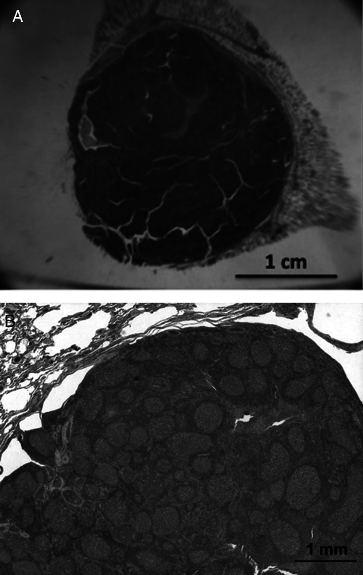
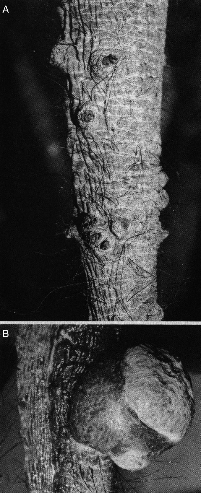
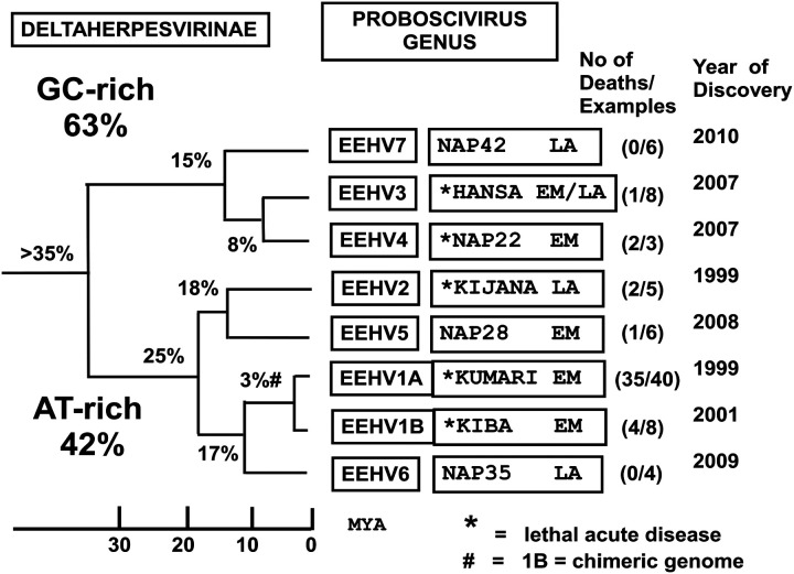
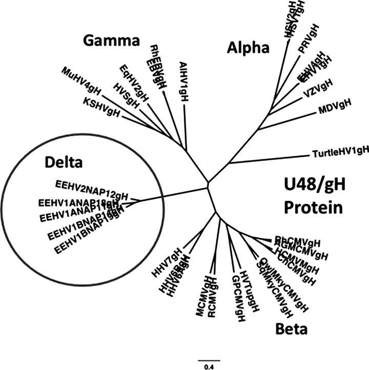
More than 100 young captive and wild Asian elephants are known to have died from a rapid-onset, acute hemorrhagic disease caused primarily by multiple distinct strains of two closely related chimeric variants of a novel herpesvirus species designated elephant endotheliotropic herpesvirus (EEHV1A and EEHV1B). These and two other species of Probosciviruses (EEHV4 and EEHV5) are evidently ancient and likely nearly ubiquitous asymptomatic infections of adult Asian elephants worldwide that are occasionally shed in trunk wash secretions. Although only a handful of similar cases have been observed in African elephants, they also have proved to harbor their own multiple and distinct species of Probosciviruses-EEHV2, EEHV3, EEHV6, and EEHV7-found in lung and skin nodules or saliva. For reasons that are not yet understood, approximately 20% of Asian elephant calves appear to be susceptible to the disease when primary infections are not controlled by normal innate cellular and humoral immune responses. Sensitive specific polymerase chain reaction (PCR) DNA blood tests have been developed, routine monitoring has been established, the complete large DNA genomes of each of the four Asian EEHV species have now been sequenced, and PCR gene subtyping has provided unambiguous evidence that this is a sporadic rather than epidemic disease that it is not being spread among zoos or other elephant housing facilities. Nevertheless, researchers have not yet been able to propagate EEHV in cell culture, determine whether or not human antiherpesvirus drugs are effective inhibitors, or develop serology assays that can distinguish between antibodies against the multiple different EEHV species.

Keywords: Elephas maximus; Loxodonta Africana; Probosciviruses; calves; elephant endotheliotropic herpesvirus (EEHV); elephant hemorrhagic disease; lung and skin nodules.


Figures

Figure 1
Figure 1
Clinical and pathological features of elephant endotheliotropic herpesvirus (EEHV)–positive elephant hemorrhagic disease. (A) Surviving 16-month-old Asian elephant calf showing early clinical symptoms of EEHV1-associated acute hemorrhagic disease including subcutaneous edema of the head, neck (white arrow), and thoracic limbs. This was the first survivor treated with FCV. (B) Gross pathological features at necropsy from a lethal Asian elephant case showing subepicardial ecchymotic hemorrhages of the heart (black arrow) attributed to EEHV1. (C) Low-power photomicrograph of typical extensive extravascular hemorrhage and separation of the myocardial fibers attributed to EEHV1. Hematoxylin and eosin, bar = 150 µm. (D) Higher-power photomicrograph of a diagnostic herpesvirus-like intranuclear inclusion body (black arrow) in a capillary endothelial cell from necropsy heart tissue. Hematoxylin and eosin, bar = 25 µm. (E) Transmission electron micrograph (TEM) of the heart from an African elephant calf that died of EEHV hemorrhagic disease demonstrating an intranuclear inclusion body (black arrow) within a capillary endothelial cell. Bar = 500 nm. (F) Higher magnification TEM of the same intranuclear inclusion body from E, showing greater detail of the unenveloped herpesvirus nucleocapsids (black arrows). Bar = 200 nm. All panels from Richman (2003).
Figure 2
Figure 2
African elephant lung nodules. Photomicrographs of follicular lymphoid nodules from culled healthy wild adult African elephants from Kruger National Park in South Africa in 1995. Two stored frozen nodules from this study in the National Elephant Herpes Laboratory collection contained very high levels of EEHV3 DNA or EEHV2 plus EEHV3 DNA. According to McCully and colleagues (1971), similar lung nodules were found in 75% of random culled African elephants in Zimbabwe. (A) Low-power photomicrographs of a large follicular lymphoid nodule found at necropsy in the lung of a wild adult Loxodonta africana. (B) Higher-power photomicrograph of a similar lung nodule showing multiple follicles composed of lymphocytes.
Figure 3
Figure 3
African elephant skin nodules. Typical herpesvirus skin nodules observed on the trunk and head of 1% of healthy juvenile African elephants in Africa, as well as in the outbreak among a large group of young orphan African calves imported from Zimbabwe to Florida (Jacobson et al. 1986). Similar skin nodules biopsied from Kenyan elephants have been found to contain DNA from multiple viruses, including EEHV2, EEHV3, EEHV7, several types of elephant gammaherpesviruses (EGHVs), and African elephant polyomavirus (V.R. Pearson, personal communication, 2013. Fox Chase Cancer center, Philadelphia; Finding EEHVs in Wild African Elephants by Virginia Pearson https://elephantmanagers.com/…/ElephantManagersArticleKWS__1_.doc. August 2012). (A) Photograph of regressed skin nodules from the trunk of an African elephant. (B) Photograph of a proliferative skin nodule from the trunk of an African elephant. Reproduced with permission from Elliot Jacobson and Journal of the American Veterinary Medical Association.
Figure 4
Figure 4
Genetic relationships among seven distinct species of elephant endotheliotropic herpesvirus (EEHV). Phylogeny-based classification of all major EEHV species within both the AT-rich and GC-rich branches showing average nucleotide divergence levels, approximate estimated ages of last common ancestors (in millions of years ago), name or case code of the prototype example, number of associated lethal cases, total number of examples of each type, and the year of first discovery.
Figure 5
Figure 5
Elephant endotheliotropic viruses (EEHVs) represent a novel subfamily of mammalian herpesviruses (circled). Radial distance-based phylogenetic tree comparing the amino-acid identity values of the intact U48(glycoprotein-H) proteins of several EEHVs with the orthologous core proteins representative of all major mammalian herpesvirus subfamilies. Because of the lack of a close match to any of the existing defined subfamilies (alpha, beta, and gamma), the EEHV clade (Proboscivirus genus) has been proposed to form a new deltaherpesvirus subfamily (Pellett 2014; Richman et al. 2014; Zong et al. 2014).
Figure 6
Figure 6
Wide range of genetic variation among elephant endotheliotropic herpesvirus (EEHV) strains. Observed amino-acid polymorphisms among polymerase chain reaction–analyzed DNA sequences for the U51(vGPCR1) protein from 22 examples of EEHV1-associated hemorrhagic disease from North American (NAP#) or European (EP#) Proboscivirus cases. The particular characteristic motif between positions 252 to 260 defines the major subtype designation (A, B, C, D, or E). * = Four cases born at the same U.S. facility.
Figure 7
Figure 7
Lack of spread of specific EEHV1 strains between elephant housing facilities and broad distribution of EEHV1 strains worldwide. Summary of the U51(vGPCR1) subtypes identified up to the year 2012 among lethal and surviving or asymptomatic elephant endotheliotropic herpesvirus (EEHV) hemorrhagic disease cases worldwide. Home and away refer, respectively, to calves born at a single facility that died at the same facility or after transfer to another facility.

References

    1. Atkins L, Zong J-C, Tan J, Mejia A, Heaggans SY, Nofs SA, Stanton JJ, Flanagan JP, Howard L, Latimer E, Stevens MR, Hoffman DS, Hayward GS, Ling PD. 2013. EEHV-5, a newly recognized elephant herpesvirus associated with clinical and subclinical infections in captive Asian elephants (Elephas maximus). J Zoo Wildl Med 44:136–143. - PMC - PubMed
    1. Bouchard B, Xaymountry B, Thongtip N, Lertwatcharasarakul P, Wajjwalku W. 2014. First reported case of elephant endotheliotropic herpes virus infection in Laos. J Zoo Wildl Med 45:704–707. - PubMed
    1. Ehlers B, Dural G, Marschall M, Schregel V, Goltz M, Hentschke J. 2006. Endotheliotropic elephant herpesvirus, the first betaherpesvirus with a thymidine kinase gene. J Gen Virol 87:2781–2789. - PubMed
    1. Fickel J, Lieckfeldt D, Richman LK, Streich WJ, Hildebrandt TB, Pitra C. 2003. Comparison of glycoprotein B (gB) variants of the elephant endotheliotropic herpesvirus (EEHV) isolated from Asian elephants (Elephas maximus). Vet Microb 91:11–21. - PubMed
    1. Fuery A, Tion J, Ling PD, Long SY, Heaggans SY, Hayward GS. 2015. EEHV infection in surviving USA Asian elephant calves. J Zoo Wildl Med In press.

Publication types

MeSH terms

LinkOut - more resources