

Cytosolic splice isoform of Hsp70 nucleotide exchange factor Fes1 is required for the degradation of misfolded proteins in yeast
- PMID: 26912797
- PMCID: PMC4831876
- DOI: 10.1091/mbc.E15-10-0697
Cytosolic splice isoform of Hsp70 nucleotide exchange factor Fes1 is required for the degradation of misfolded proteins in yeast
Abstract
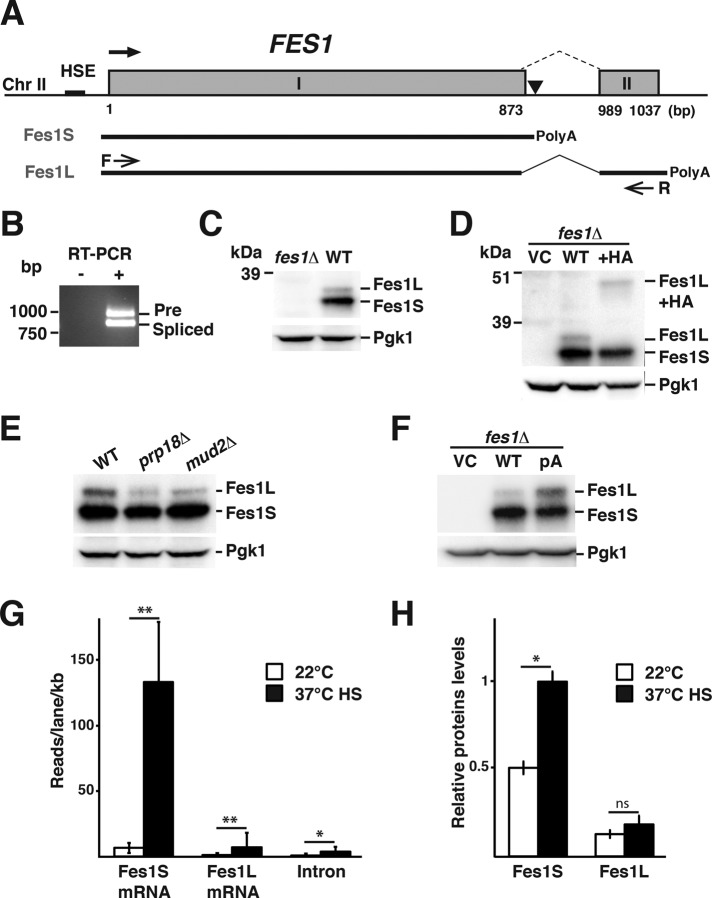
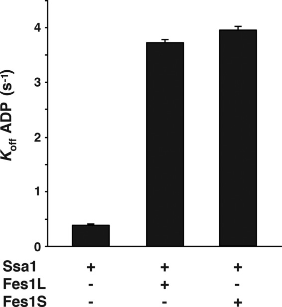
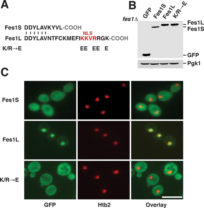
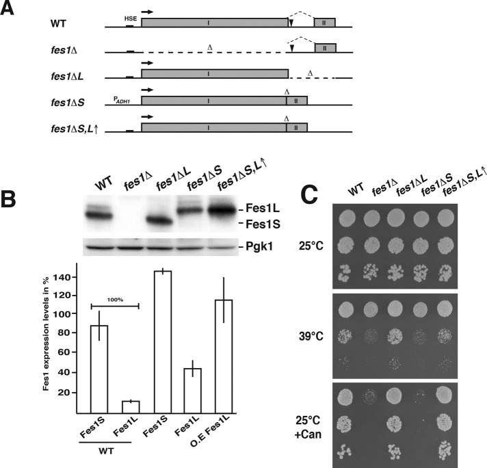
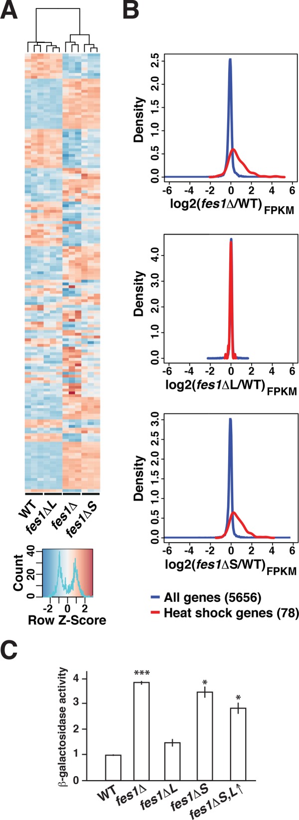
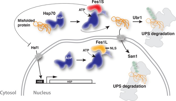
Cells maintain proteostasis by selectively recognizing and targeting misfolded proteins for degradation. InSaccharomyces cerevisiae, the Hsp70 nucleotide exchange factor Fes1 is essential for the degradation of chaperone-associated misfolded proteins by the ubiquitin-proteasome system. Here we show that theFES1transcript undergoes unique 3' alternative splicing that results in two equally active isoforms with alternative C-termini, Fes1L and Fes1S. Fes1L is actively targeted to the nucleus and represents the first identified nuclear Hsp70 nucleotide exchange factor. In contrast, Fes1S localizes to the cytosol and is essential to maintain proteostasis. In the absence of Fes1S, the heat-shock response is constitutively induced at normally nonstressful conditions. Moreover, cells display severe growth defects when elevated temperatures, amino acid analogues, or the ectopic expression of misfolded proteins, induce protein misfolding. Importantly, misfolded proteins are not targeted for degradation by the ubiquitin-proteasome system. These observations support the notion that cytosolic Fes1S maintains proteostasis by supporting the removal of toxic misfolded proteins by proteasomal degradation. This study provides key findings for the understanding of the organization of protein quality control mechanisms in the cytosol and nucleus.
© 2016 Gowda et al. This article is distributed by The American Society for Cell Biology under license from the author(s). Two months after publication it is available to the public under an Attribution–Noncommercial–Share Alike 3.0 Unported Creative Commons License (http://creativecommons.org/licenses/by-nc-sa/3.0).
Figures

Similar articles
-
Hsp70 nucleotide exchange factor Fes1 is essential for ubiquitin-dependent degradation of misfolded cytosolic proteins.Proc Natl Acad Sci U S A. 2013 Apr 9;110(15):5975-80. doi: 10.1073/pnas.1216778110. Epub 2013 Mar 25. Proc Natl Acad Sci U S A. 2013. PMID: 23530227 Free PMC article.
-
The Type II Hsp40 Sis1 cooperates with Hsp70 and the E3 ligase Ubr1 to promote degradation of terminally misfolded cytosolic protein.PLoS One. 2013;8(1):e52099. doi: 10.1371/journal.pone.0052099. Epub 2013 Jan 16. PLoS One. 2013. PMID: 23341891 Free PMC article.
-
Hierarchical functional specificity of cytosolic heat shock protein 70 (Hsp70) nucleotide exchange factors in yeast.J Biol Chem. 2014 May 9;289(19):13155-67. doi: 10.1074/jbc.M113.530014. Epub 2014 Mar 26. J Biol Chem. 2014. PMID: 24671421 Free PMC article.
-
Co-Chaperones in Targeting and Delivery of Misfolded Proteins to the 26S Proteasome.Biomolecules. 2020 Aug 4;10(8):1141. doi: 10.3390/biom10081141. Biomolecules. 2020. PMID: 32759676 Free PMC article. Review.
-
Spatially organized aggregation of misfolded proteins as cellular stress defense strategy.J Mol Biol. 2015 Apr 10;427(7):1564-74. doi: 10.1016/j.jmb.2015.02.006. Epub 2015 Feb 11. J Mol Biol. 2015. PMID: 25681695 Review.
Cited by
-
Cellular sequestrases maintain basal Hsp70 capacity ensuring balanced proteostasis.Nat Commun. 2019 Oct 24;10(1):4851. doi: 10.1038/s41467-019-12868-1. Nat Commun. 2019. PMID: 31649258 Free PMC article.
-
OsFes1C, a potential nucleotide exchange factor for OsBiP1, is involved in the ER and salt stress responses.Plant Physiol. 2021 Sep 4;187(1):396-408. doi: 10.1093/plphys/kiab263. Plant Physiol. 2021. PMID: 34618140 Free PMC article.
-
Hsp40/70/110 chaperones adapt nuclear protein quality control to serve cytosolic clients.J Cell Biol. 2018 Jun 4;217(6):2019-2032. doi: 10.1083/jcb.201706091. Epub 2018 Apr 13. J Cell Biol. 2018. PMID: 29653997 Free PMC article.
-
BRF Negatively Regulates Thermotolerance Defect of fes1a in Arabidopsis.Front Plant Sci. 2020 Mar 10;11:171. doi: 10.3389/fpls.2020.00171. eCollection 2020. Front Plant Sci. 2020. PMID: 32210987 Free PMC article.
-
Acquired Resistance to Severe Ethanol Stress in Saccharomyces cerevisiae Protein Quality Control.Appl Environ Microbiol. 2021 Feb 26;87(6):e02353-20. doi: 10.1128/AEM.02353-20. Print 2021 Feb 26. Appl Environ Microbiol. 2021. PMID: 33361368 Free PMC article.
References
-
- Abovich N, Liao XC, Rosbash M. The yeast MUD2 protein: an interaction with PRP11 defines a bridge between commitment complexes and U2 snRNP addition. Genes Dev. 1994;8:843–854. - PubMed
-
- Andréasson C, Fiaux J, Rampelt H, Mayer MP, Bukau B. Hsp110 is a nucleotide-activated exchange factor for Hsp70. J Biol Chem. 2008b;283:8877–8884. - PubMed
Publication types
MeSH terms
Substances
LinkOut - more resources
Full Text Sources
Other Literature Sources
Molecular Biology Databases
