. 2016 Feb 12;351(6274):711-4.
doi: 10.1126/science.aad2791.

Pathogenic CD4 T cells in type 1 diabetes recognize epitopes formed by peptide fusion

Affiliations

Pathogenic CD4 T cells in type 1 diabetes recognize epitopes formed by peptide fusion

Thomas Delong et al. Science. .

Abstract

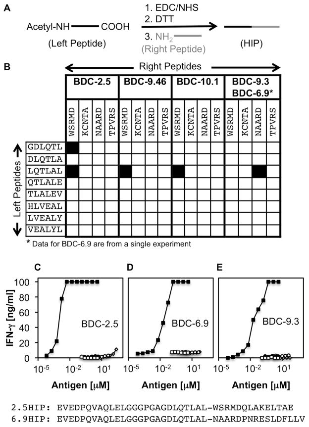
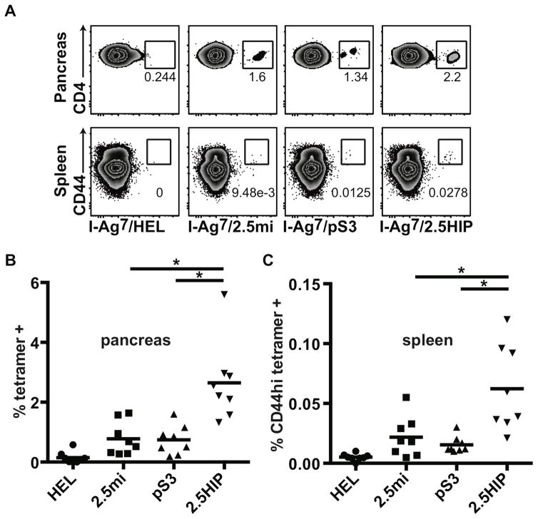
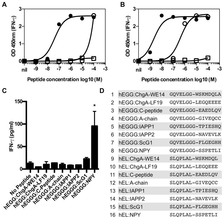
T cell-mediated destruction of insulin-producing β cells in the pancreas causes type 1 diabetes (T1D). CD4 T cell responses play a central role in β cell destruction, but the identity of the epitopes recognized by pathogenic CD4 T cells remains unknown. We found that diabetes-inducing CD4 T cell clones isolated from nonobese diabetic mice recognize epitopes formed by covalent cross-linking of proinsulin peptides to other peptides present in β cell secretory granules. These hybrid insulin peptides (HIPs) are antigenic for CD4 T cells and can be detected by mass spectrometry in β cells. CD4 T cells from the residual pancreatic islets of two organ donors who had T1D also recognize HIPs. Autoreactive T cells targeting hybrid peptides may explain how immune tolerance is broken in T1D.


Figures

Fig. 1
Fig. 1. Identification of HIP sequences that are recognized by pathogenic T cell clones
(A) HIPs were synthesized upon chemical activation of the C-termini of acetylated left peptides with EDC/NHS, followed by quenching of residual EDC with DTT. Addition of a right peptide leads to covalent linkage of the right peptide N-terminal amine to the activated C-terminus of the left peptide. (B) A library of 32 HIPs was synthesized. Left peptides are C-terminal amino acid sequences of various mouse insulin C-peptide and B-Chain fragments. Right peptides reflect the N-terminal amino acid sequences of mouse WE14 (WSRMD), IAPP1 (TPVRS), Amylin (KCNTA) and IAPP2 (NAARD). T cell clones BDC-2.5, BDC-10.1, BDC-9.46 and BDC-9.3 were used to screen the peptide library. Black squares indicate positive T cell responses to individual HIPs. Data shown are from three separate experiments. To confirm antigenicity of HIPs, pure peptides (>95%) were obtained commercially for assay with the T cell clones: (C) WE14-reactive clones such as BDC-2.5 respond to 2.5HIP (solid squares) in the low nanomolar range whereas they are stimulated by unmodified WE14 only at high concentrations (100 μM) (open diamonds). IAPP-reactive clones, BDC-6.9 (D) and BDC-9.3 (E), respond to low nanomolar concentrations of the 6.9HIP (solid squares), but were not responsive to IAPP2 alone (open diamonds). None of the clones responds to Insulin 2 C-peptide (open circles) or the Insulin 2 C-peptide fragment ending with the amino acid sequence DLQTLAL (solid circles). Co-incubating the C-Peptide fragment with WE14 or IAPP2 without covalent linkage (open squares) did not increase antigenicity. Amino acid sequences for 2.5HIP and 6.9HIP are shown at the bottom of Fig. 1. Data are representative of at least three separate experiments.
Fig. 2
Fig. 2. Identification of HIPs in antigenic β-cell fractions
(A) Size exclusion chromatography (SEC) fractions highly enriched for antigen were further fractionated by RP-HPLC (black line). IFN-γ T cell responses to individual fractions are shown for BDC-2.5 (grey line). (B) Following the proteolytic digest with AspN, the targeted MS/MS analysis of antigen-containing fractions reveals the spectrum of the HIP that contains the C-peptide amino acid sequence DLQTLAL and the WE14 sequence WSRM. (C) The corresponding 6.9HIP (DLQTLAL-NAAR) could also be identified in AspN digested fractions that contain the antigen recognized by BDC-6.9 and BDC-9.3. Data for chromatographic fractionation are representative of at least three separate experiments. Spectra shown are representative and were obtained from two separate experiments.
Fig. 3
Fig. 3. Tetramer analysis of pancreas and spleen of diabetic NOD mice
Single cell suspensions were prepared from pancreas and spleen of diabetic NOD female mice (n = 8) and stained with tetramers, antibodies and a live cell marker (7AAD). Gates were set on live leukocytes (7AAD-, CD45), CD4, dump- (CD8, CD11b, CD11c, CD19, F4/80) cells. (A) Tetramer staining in the pancreas and the spleen of a representative mouse. (B) Summary of CD4 tetramer-positive cells present in the pancreas. (C) Summary of CD4 CD44hi tetramer-positive cells present in the spleen. Each symbol represents a different mouse. Averages are indicated as a black horizontal bar. Data are from 4 independent experiments and were analyzed by two-tailed unpaired t-test. Statistical significance (*) was defined at a p value <0.05.
Fig. 4
Fig. 4. Human islet-infiltrating T cells respond to HIPs
Human islet-infiltrating CD4 T cell clones A3.10 (A), A2.11 (B) and the cell line MG1 (C) respond to HIPs. (A and B) Clones from isolated islets of a 19 year-old male with 3 years of T1D were incubated with the indicated concentration of peptide and HLA-DQ2+, DQ8+ EBV transformed B-cell line overnight. Responses to peptide were detected by IFN-γ secretion measured by ELISA. Each point represents the mean and standard deviation of triplicate wells. Solid circles are the HIP (GQVELGGG-NAVEVLK), open circles are proinsulin 37–54 (LQVELGGGPGAGSLQ) open squares are another HIP incorporating the same region of IAPP2 (SLQPLAL-NAVEVLK). Clone A3.10 and A2.11 (Fig. S6) recognize HIPs presented by HLA-DQ8. (C) A CD4 T cell line grown from isolated islets of a 20 year-old male with 7 years of T1D (DR17, DR4, DQ2, DQ8) secreted IFN-γ when a HIP, formed by the fusion of proinsulin C-peptide to the pancreatic polypeptide, neuropeptide Y (GQVELGGG-SSPETLI), was presented by Priess B cells (DR4+/+, DQ8+/+); all peptides at 20μg/ml. *p=0.0395 as compared to the media control (paired Student’s t test). The mean and standard deviation of triplicates in shown in all graphs. For all data, one representative of at least two independent experiments is shown. (D) List of HIPs (>95% purity) screened.

References

    1. Atkinson MA, Leiter EH. The NOD mouse model of type 1 diabetes: as good as it gets? Nature medicine. 1999 Jun;5:601. - PubMed
    1. Nakayama M, et al. Prime role for an insulin epitope in the development of type 1 diabetes in NOD mice. Nature. 2005 May 12;435:220. - PMC - PubMed
    1. Haskins K. Pathogenic T-cell clones in autoimmune diabetes: more lessons from the NOD mouse. Advances in immunology. 2005;87:123. - PubMed
    1. Stadinski BD, et al. Chromogranin A is an autoantigen in type 1 diabetes. Nature immunology. 2010 Mar;11:225. - PMC - PubMed
    1. Delong T, et al. Islet amyloid polypeptide is a target antigen for diabetogenic CD4+ T cells. Diabetes. 2011 Sep;60:2325. - PMC - PubMed

Publication types

MeSH terms