Review
. 2016 Feb 15:7:36.
doi: 10.3389/fimmu.2016.00036. eCollection 2016.

Early Components of the Complement Classical Activation Pathway in Human Systemic Autoimmune Diseases

Affiliations
Review

Early Components of the Complement Classical Activation Pathway in Human Systemic Autoimmune Diseases

Katherine E Lintner et al. Front Immunol. .

Abstract

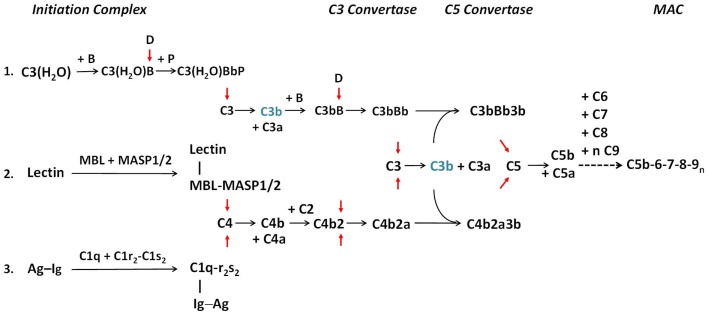
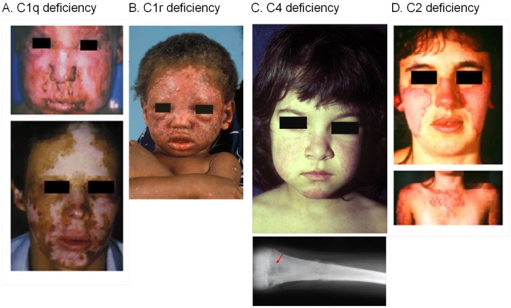
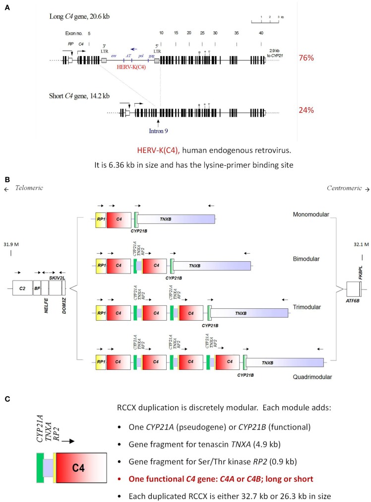
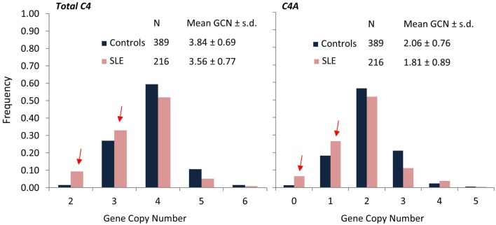
The complement system consists of effector proteins, regulators, and receptors that participate in host defense against pathogens. Activation of the complement system, via the classical pathway (CP), has long been recognized in immune complex-mediated tissue injury, most notably systemic lupus erythematosus (SLE). Paradoxically, a complete deficiency of an early component of the CP, as evidenced by homozygous genetic deficiencies reported in human, are strongly associated with the risk of developing SLE or a lupus-like disease. Similarly, isotype deficiency attributable to a gene copy-number (GCN) variation and/or the presence of autoantibodies directed against a CP component or a regulatory protein that result in an acquired deficiency are relatively common in SLE patients. Applying accurate assay methodologies with rigorous data validations, low GCNs of total C4, and heterozygous and homozygous deficiencies of C4A have been shown as medium to large effect size risk factors, while high copy numbers of total C4 or C4A as prevalent protective factors, of European and East-Asian SLE. Here, we summarize the current knowledge related to genetic deficiency and insufficiency, and acquired protein deficiencies for C1q, C1r, C1s, C4A/C4B, and C2 in disease pathogenesis and prognosis of SLE, and, briefly, for other systemic autoimmune diseases. As the complement system is increasingly found to be associated with autoimmune diseases and immune-mediated diseases, it has become an attractive therapeutic target. We highlight the recent developments and offer a balanced perspective concerning future investigations and therapeutic applications with a focus on early components of the CP in human systemic autoimmune diseases.

Keywords: autoimmune diseases; classical pathway; complement C1q; complement C1r; complement C1s; complement C2; complement C4; systemic lupus erythematosus.


Figures

Figure 1
Figure 1
The activation pathways of the complement system. The three activation pathways of the complement system are shown according to evolution and physiologic sequences. Pathway 1 is known as the alternative pathway. It is activated through a tick-over mechanism because of continuous hydrolysis of the thioester bond in C3, which enables the formation with factor B to form a weak C3 convertase. Pathway 2 is known as the MBL or lectin pathway. It is initiated through the binding of mannan-binding lectin (MBL) or ficolin to arrays of simple sugar molecules in glycosylated antigens on microbes. This is a pattern recognition mechanism characteristic of the innate immune system. Pathway 3 is known as the classical pathway and is initiated through the binding of specific antibodies IgM or IgG to antigens. It is an effector arm of the humoral adaptive immune system. Each activation pathway engages the formation of a multi-molecular initiation complex, followed by the assembly of a C3 convertase and a C5 convertase for activation of C3 and C5, respectively and culminates in the formation of membrane attack complexes (MAC) on the target membrane. All three pathways can be amplified through a positive feedback mechanism, as C3b (in blue) generated by any C3 convertase can feed to the alternative pathway through association with factor B to form new C3 convertase after activation by factor D (pathway 1). Anaphylatoxins C3a and C5a are produced during the activation process. For brevity, by-products generated during the activation of C2 and factor B are not shown. Red arrows show activation of component proteins through cleavage by serine proteases. A dotted horizontal arrow denotes multiple steps are involved in the formation of the membrane attack complex. Early components of the classical pathway C1q, C1r, C1s, and C4 are engaged in the differentiation of immunity and autoimmunity, as genetic or acquired deficiency in any of these components are linked to pathogenesis of SLE. Complement C2 is also involved in the protection against autoimmunity but its effect size is smaller [modified from Ref. (2)].
Figure 2
Figure 2
Typical serial serum protein profiles of complement C4 and C3 in human SLE patients. Serum C4 (red, solid line) and C3 (green, dashed line) protein levels tend to go up and down together in most SLE patients. The horizontal dotted line indicates the low level of serum C4 (<10 mg/dL), below which usually requires clinical attention. The profiles shown are taken from three individual patients over a 24-month period and represent three common profiles typically observed in SLE patients. In the first profile (A), levels of C4 and C3 were chronically low. In some patients, even if C3 levels rose to normal range, C4 levels remained low. Patients with this profile are often characterized by low copy-number of C4 genes. (B) The second profile had frequent and parallel fluctuations of serum C3 and C4. Patients with this profile had active disease, and low C3 and low C4 roughly correlated with disease activity. In the third profile (C), C4 and C3 protein levels stayed in the normal range most of the time, except at the time of diagnosis and during a disease relapse. Patients with this profile had relatively inactive disease. Patients with the second and third profiles have normal gene copy-number of total C4 but may have a heterozygous deficiency of C4A [modified from Reference (12)].
Figure 3
Figure 3
SLE patients with a homozygous deficiency of early components for the classical pathway of complement activation. Severe cutaneous lesions are common clinical presentations in SLE patients with a complete complement deficiency. (A) A homozygous C1q-deficient male child with cutaneous infection (upper panel) and with discoid lupus erythematosus and scarring lesions on face when he was 22 years old (lower panel). (B) A male child with discoid lupus at 16-month old with homozygous C1r-deficiency. This patient experienced generalized seizures, developed a scissoring gait with toe walking, spasticity and weakness of the legs. At 18 years old, he was diagnosed with class IV lupus nephritis and progressed to end-stage renal disease. (C) A complete C4-deficient girl at 3 years old with butterfly rash and cheilitis (upper panel), and osteomyelitis of the femur at 10 years old (lower panel). This patient died at age 12 because of pulmonary infection and cardiovascular failure. (D) A homozygous C2-deficient young woman with acute cutaneous lupus erythematosus. The upper panel shows the butterfly rash, and the lower panel shows photosensitive lesions on sun-exposed areas [adopted from Ref. (–43)].
Figure 4
Figure 4
Gene size dichotomy and gene copy-number variation of complement C4. A human C4 gene consists of 41 exons coding for a precursor protein of 1744 amino acids including a signal peptide of 19 amino acids. (A) There are two forms of C4 genes. The long gene is 20.6 kb, and the short gene is 14.2 kb. In a long C4 gene, an endogenous retrovirus HERV-K (C4), which is 6.4 kb in size, integrated into its ninth intron. Among healthy subjects of European ancestry, 76% of C4 genes belong to the long form and 24% belong to the short form. (B) Among European subjects, one to four copies of C4 genes are present in the central region of the major histocompatibility complex (MHC) located on chromosome 6p21.3. Thus, there is a continuous variation in copy number of C4 genes from two to eight copies among different human subjects. (C) The duplication of a C4 gene occurs in a modular fashion, with a 0.9 kb fragment of RP (STK19) upstream of complement C4, a full steroid cytochrome P450 21-hydroxylase (CYP21) and a 4.0 kb fragment of the tenascin (TNX) at the downstream region of C4 (known as a RCCX module). The duplication of CYP21 gene can be a pseudogene (CYP21A or CYP21A1P) or an intact functional gene (CYP21B or CYP21A2). Each C4 gene in the RCCX module may either code for an acidic C4A or a basic C4B. Each C4 gene may be either long or short [adopted from Ref. (–99)].
Figure 5
Figure 5
Comparisons of frequencies for total C4, C4A, and C4B gene copy-number groups in SLE (red) and controls (blue). SLE patients (N = 216) of European ancestry showed significantly higher frequencies for lower copy-numbers of total C4 (GCN = 2 or 3) and C4A (GCN = 0 or 1) compared to healthy, race-matched controls (N = 389). Mean GCN for Total C4 in SLE (3.56 ± 0.77) was significantly lower than in controls (3.84 ± 0.69; p = 5.3 × 10−6, t-test). Similarly, mean GCN for C4A in SLE (1.81 ± 0.89) was significantly lower than in controls (2.06 ± 0.76; p = 2.0 × 10−4, t-test) [modified from Ref. (13)].
Figure 6
Figure 6
Race-specific distribution patterns of RCCX modules in human populations. The size dichotomy of C4 genes and copy-number variation of RCCX modules on an MHC haplotype together create a repertoire of length variants among different human subjects, which also exist with race-specific distribution patterns. (A) The most prevalent haplotypes of RCCX in Whites and Asian-Indians are the bimodular long-long (LL) and bimodular long-short (LS) in Blacks and East-Asians. (B) Notably, monomodular-short (mono-S or S) haplotypes with a single short C4B gene and C4A deficiency is relatively common in White and Black subjects but almost absent in Asians [modified from Ref. (118)].

References

    1. Ricklin D, Hajishengallis G, Yang K, Lambris JD. Complement: a key system for immune surveillance and homeostasis. Nat Immunol (2010) 11(9):785–97.10.1038/ni.1923 - DOI - PMC - PubMed
    1. Yu C, Driest K, Wu Y, Lintner K, Patwardhan A, Spencer CH, et al. Complement in rheumatic diseases. In: Diamond B, Davidson A, editors. Encyclopedia of Medical Immunology: Autoimmune Diseases. New York, NY: Springer; (2014). p. 286–302.
    1. Atkinson J, Yu C. The complement system in systemic lupus erythematosus. In: Tsokos GC, editor. Systemic Lupus Erythematosus. Chapter 12, New York, NY: Elsevier; (2016). p. 81–112.10.1016/B978-0-12-801917-7.00012-7 - DOI
    1. Sturfelt G, Truedsson L. Complement in the immunopathogenesis of rheumatic disease. Nat Rev Rheumatol (2012) 8(8):458–68.10.1038/nrrheum.2012.75 - DOI - PubMed
    1. Pillemer L, Blum L, Lepow IH, Ross OA, Todd EW, Wardlaw AC. The properdin system and immunity. I. Demonstration and isolation of a new serum protein, properdin, and its role in immune phenomena. Science (1954) 120(3112):279–85.10.1126/science.120.3112.279 - DOI - PubMed