Review
. 2015 Aug 25;25(4):221-30.
doi: 10.4081/ejtm.2015.5272. eCollection 2015 Aug 24.

Biology of Muscle Atrophy and of its Recovery by FES in Aging and Mobility Impairments: Roots and By-Products

Affiliations
Review

Biology of Muscle Atrophy and of its Recovery by FES in Aging and Mobility Impairments: Roots and By-Products

Ugo Carraro et al. Eur J Transl Myol. .

Abstract

There is something in our genome that dictates life expectancy and there is nothing that can be done to avoid this; indeed, there is not yet any record of a person who has cheated death. Our physical prowess can vacillate substantially in our lifetime according to our activity levels and nutritional status and we may fight aging, but we will inevitably lose. We have presented strong evidence that the atrophy which accompanies aging is to some extent caused by loss of innervation. We compared muscle biopsies of sedentary seniors to those of life long active seniors, and show that these groups indeed have a different distribution of muscle fiber diameter and fiber type. The senior sportsmen have many more slow fiber-type groupings than the sedentary people which provides strong evidence of denervation-reinnervation events in muscle fibers. It appears that activity maintains the motoneurons and the muscle fibers. Premature or accelerated aging of muscle may occur as the result of many chronic diseases. One extreme case is provided by irreversible damage of the Conus and Cauda Equina, a spinal cord injury (SCI) sequela in which the human leg muscles may be completely and permanently disconnected from the nervous system with the almost complete disappearance of muscle fibers within 3-5 years from SCI. In cases of this extreme example of muscle degeneration, we have used 2D Muscle Color CT to gather data supporting the idea that electrical stimulation of denervated muscles can retain and even regain muscle. We show here that, if people are compliant, atrophy can be reversed. A further example of activity-related muscle adaptation is provided by the fact that mitochondrial distribution and density are significantly changed by functional electrical stimulation in horse muscle biopsies relative to those not receiving treatment. All together, the data indicate that FES is a good way to modify behaviors of muscle fibers by increasing the contraction load per day. Indeed, it should be possible to defer the muscle decline that occurs in aging people and in those who have become unable to participate in physical activities. Thus, FES should be considered for use in rehabilitation centers, nursing facilities and in critical care units when patients are completely inactive even for short periods of time.

Keywords: Muscle power; aging decay; equine muscle spasm; h-b FES-induced muscle recovery; long-term denervated muscles; master athletes; muscle denervation/reinnervation; subsarcolemmal mitochondria; type groupings.


Figures

Fig. 1.
Fig. 1.
Age-related decline of skeletal muscle power derived from world records of running, jumping and throwing events of Masters of different age classes. Lines: Light blue, 100 meters run; Red, 400 meters run; Blue, Long Jump; Gray, High Jump; Yellow, Shot Put; Green, Hammer Throw. The insets show Usain Bolt (around 30 years), Paolo Gava (< 60 years) and a 90 year old Japanese Master world record-holding athlete pictured the day they established the respective world record. Whatever the extent of training, even in the extreme cases of Master world record-holding men, muscle power almost linearly decreases with age pointing to around 110 years of human survival. The points of each individual line represent the normalized power of each World Record of the Master Athletes (seldom, the same athlete was able to hold the record for different age classes). The size changes of muscles and of myofibers do not fully explain the extent of dysfunction observed during aging. Though muscle disorders of ultrastructure and of molecular mechanisms may explain the additional functional decline, we are interested in studying if muscle fiber atrophy/apoptosis driven by denervation is a contributing factor (see Fig. 2).
Fig. 2.
Fig. 2.
In muscle biopsies of lifelong highly active senior amateur sportsmen, the MHC co-expression in type-grouped slow muscle fibers suggests that activity-driven reinnervation by preferentially saved slow type motoneurons occurs. The MHC co-expressing myofibers are of normal size and some of these fill the gaps between clusters of slow myofibers (that is, slow type-groupings). We suggest these fibers were denervated fast muscle fibers preferentially reinnervated by axons sprouting from slow motoneurons.
Fig. 3.
Fig. 3.
Permanent long-term denervation simulates premature aging in muscle. Human skeletal muscle undergoes four defined phases subsequent to permanent long-term denervation: 1) Loss of contractlity and ultrastructural disorganization (in months); 2) Atrophy (up to 2-years after SCI); 3) Severe atrophy (3 to 6 ys after SCI); 4) Loss of myofibers and muscle degeneration (> 3 years after SCI). These are the unexpected results of the EU Program RISE: Use of electrical stimulation to restore standing in paraplegics with long-term denervated degenerated muscles (Contract no. QLG5-CT-2001-02191).
Fig. 4.
Fig. 4.
Recovery from permanent denervation (i.e., premature muscle aging) by h-b FES: the 2D Color CT evidence. Color scans of thigh muscles before (B to E) and after 2 years (G to J) of home-based functional electrical stimulation (h-b FES). Each panel shows that the cross-sectional area and the quality of quadriceps muscles in patients starting h-b FES at different time points after denervation (B, 1.2; C, 1.7; D, 3.2; E, 5.4 years) increased after 2 years of home training (G, H, I, J, respectively). Moreover, the interstitial tissues that increase with the denervation time (compare yellow, green, and blue areas in panels B, C, D, and E) decreased in the respective patient after 2 years of h-b FES (G, H, I, J, respectively).
Fig. 5.
Fig. 5.
3 D Color Muscle CT reconstruction of the rectus femoris. Reversible h-b FES-induced recovery The patient started h-b FES in 2003 (depicted in yellow). After 5 years of FES the muscle increased in size and density (2008). After 5 additional years without h-b FES the muscle appears, as would be expected, for a non-stimulated denervated muscles, i.e., even more atrophic and fibrotic (2013). The mean muscle density (expressed in Hounsfield Unit, HU) at different times from SCI are indicated by the ascending and descending light blue line.
Fig. 6.
Fig. 6.
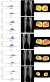
2D muscle color CT scan. The distribution analysis of the ranges of Hounsfield Unit in the histograms (left panels) allows for much more detailed quantitation of the changes occurring within the soft tissues of the leg (i.e., fat in yellow, muscles in red or orange, according to their density). During inpatient 2 months rehabilitation, the subject was treated each day for 30 minutes with electrical stimulation for denervated muscles, bilaterally on the anterior and lateral side of the leg. Histograms of HU distribution show that the right leg contains the same amounts of subcutaneous fat as the left leg, but more intramuscular fat and low density muscle at the expense of the normal density muscle. Two months of conventional physiotherapy and electrical stimulation improved in the right leg by + 5 % the content of low density muscle at the expenses of the intramuscular fat and fibrous-dense connective tissue (results not presented).
Fig. 7.
Fig. 7.
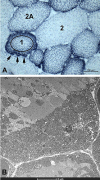
Mitochondrial evidence of FES effectiveness. A. NADH-TR stain. Type 2 large, glycolytic muscle fiber with low-density mitochondria, Type 2A medium, glycolytic-oxidative muscle fiber, Type 1 small, oxidative muscle fiber with high-density mitochondria patches (black arrows). The circle defines the central intermyofibrillar area from the coronal subsarcolemmal high-density mitochondrial area. B. Electron microscopy of high density mitochondrial patches. Note that the patches are located between capillaries (upper right and lower left corners), i.e., in paravascular location.

References

    1. Hill AV. The physiological basis of athletic records. The Scientific Monthly 1925;2:409–428.
    1. Mitchell WK, Williams J, Atherton P, Larvin M, Lund J, Narici M. Sarcopenia, dynapenia, and the impact of advancing age on human skeletal muscle size and strength; a quantitative review. Front Physiol 2012. Jul 11;3:260 doi: 10.3389/fphys.2012.00260. eCollection 2012. - PMC - PubMed
    1. Gava P, Kern H, Carraro U. Age-associated power decline from running, jumping, and throwing male masters world records. Exp Aging Res. 2015;41(2):115–35. doi: 10.1080/0361073X.2015.1001648. - PubMed
    1. Mosole S, Rossini K, Kern H, et al. Significant increase of vastus lateralis reinnervation in 70-year sportsmen with a lifelong history of high-level exercise. Eur J Transl Myol - Basic Appl Myol 2013;23:117–22. - PMC - PubMed
    1. Mosole S, Carraro U, Kern H, et al. Long-term high-level exercise promotes muscle reinnervation with age. J Neuropathol Exp Neurol 2014;73:284–94. doi: 10.1097/NEN.0000000000 000032. - PubMed