

Ultrastructural and Morphalogical Changes of Mouse Ovarian Tissues Following Direct Cover Vitrification with Different Cryoprotectants
- PMID: 26913232
- PMCID: PMC4508352
Ultrastructural and Morphalogical Changes of Mouse Ovarian Tissues Following Direct Cover Vitrification with Different Cryoprotectants
Abstract
Background: Cryopreservation of mammalian ovaries has been reported with different levels of success. Cryopreservation of ovarian tissue may be a potential alternative for treatment of infertility and many attempts have been done to improve the efficiency of ovarian cryopreservation. The objective of the present study was to compare the direct cover vitrification (DCV) with ethylene glycol (EG), dimethyl sulfoxide (DMSO) and EG plus DMSO.
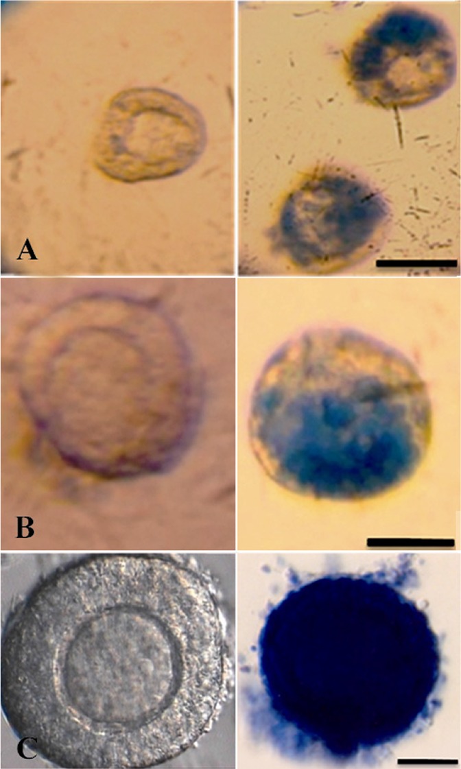
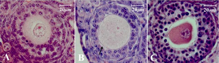
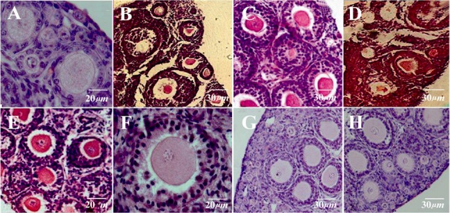
Methods: Eighty five mice were sacrificed by cervical dislocation and their ovaries were cryopreserved in the presence of 5% EG or DMSO alone or as mixture, 10% EG or DMSO alone or as mixture and a group with ascending concentrations of cryoprotectants. After toxicity testing and vitrification warming, the ovaries were fixed for histological and ultrastructural studies. In addition, the viability of mechanically isolated follicles was studied by trypan blue staining. All data were compared by ANOVA (p<0.05).
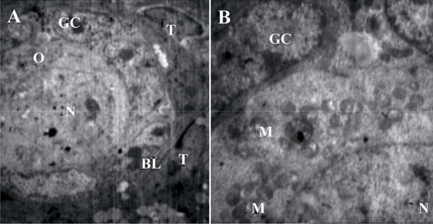
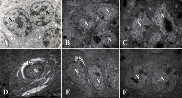
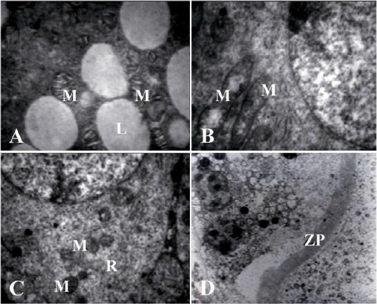
Results: Ovarian tissues frozen in EG plus DMSO in ascending concentrations retained a higher percentage of morphologically normal and or viable follicles than tissues frozen in 10 M EG plus DMSO or in either concentration of EG and DMSO alone (p<0.001). Ultrastructural analysis of ovarian tissues frozen in ascending concentrations of EG plus DMSO showed that these follicles were well preserved and it was very similar to the control group.
Conclusion: Cryopreservation of ovarian tissue in EG plus DMSO is the most effective method for preserving the structural integrity of follicles within the ovary.
Keywords: Cryopreservation; Direct cover vitrification; Ovarian tissue.
Figures
References
-
- Kuleshova LL, Lopata A. Vitrification can be more favorable than slow cooling. Fertil Steril. 2002; 78 ( 3): 449– 54. - PubMed
-
- Courbiere B, Odagescu V, Baudot A, Massardier J, Mazoyer C, Salle B, et al. Cryopreservation of the ovary by vitrification as an alternative to slow-cooling protocols. Fertil Steril. 2006; 86 ( 4 Suppl): 1243– 51. - PubMed
-
- Li YB, Zhou CQ, Yang GF, Wang Q, Dong Y. Modified vitrification method for cryopreservation of human ovarian tissues. Chin Med J. 2007; 120 ( 2): 110– 4. - PubMed
-
- Hani T, Tachibe T, Shingai S, Kamada N, Ueda O, Jishage K. Fertility of mice receiving vitrified adult mouse ovaries. Reproduction. 2006; 131 ( 4): 681– 7. - PubMed
-
- Hasegawa A, Mochida N, Ogasawara T, Koyama K. Pup birth from mouse oocytes in preantral follicles derived from vitrified and warmed ovaries followed by in vitro growth, in vitro maturation, and in vitro fertilization. Fertil Steril. 2006; 86 ( 4 Suppl): 1182– 92. - PubMed
LinkOut - more resources
Full Text Sources