

Myelin Water Fraction Is Transiently Reduced after a Single Mild Traumatic Brain Injury--A Prospective Cohort Study in Collegiate Hockey Players
- PMID: 26913900
- PMCID: PMC4767387
- DOI: 10.1371/journal.pone.0150215
Myelin Water Fraction Is Transiently Reduced after a Single Mild Traumatic Brain Injury--A Prospective Cohort Study in Collegiate Hockey Players
Abstract
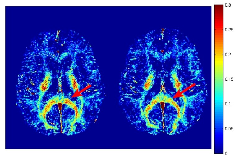
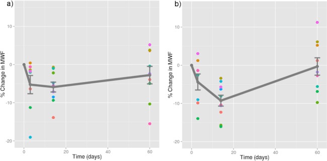
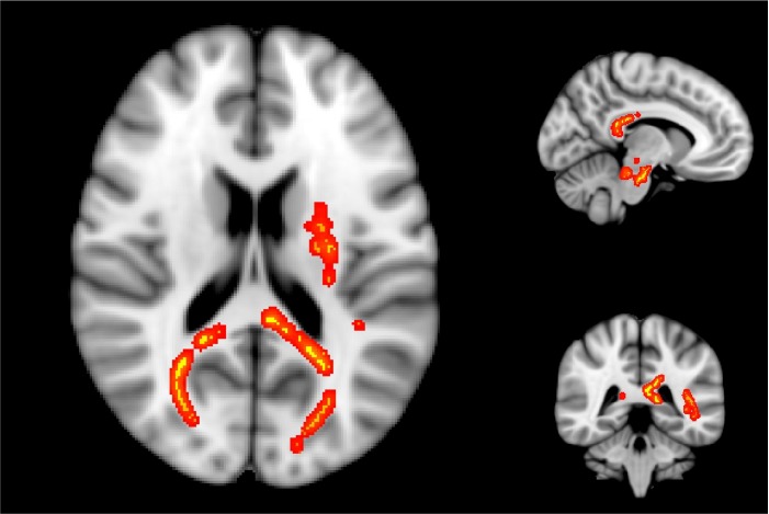
Impact-related mild traumatic brain injuries (mTBI) are a major public health concern, and remain as one of the most poorly understood injuries in the field of neuroscience. Currently, the diagnosis and management of such injuries are based largely on patient-reported symptoms. An improved understanding of the underlying pathophysiology of mTBI is urgently needed in order to develop better diagnostic and management protocols. Specifically, dynamic post-injury changes to the myelin sheath in the human brain have not been examined, despite 'compromised white matter integrity' often being described as a consequence of mTBI. In this preliminary cohort study, myelin water imaging was used to prospectively evaluate changes in myelin water fraction, derived from the T2 decay signal, in two varsity hockey teams (45 players) over one season of athletic competition. 11 players sustained a concussion during competition, and were scanned at 72 hours, 2 weeks, and 2 months post-injury. Results demonstrated a reduction in myelin water fraction at 2 weeks post-injury in several brain areas relative to preseason scans, including the splenium of the corpus callosum, right posterior thalamic radiation, left superior corona radiata, left superior longitudinal fasciculus, and left posterior limb of the internal capsule. Myelin water fraction recovered to pre-season values by 2 months post-injury. These results may indicate transient myelin disruption following a single mTBI, with subsequent remyelination of affected neurons. Myelin disruption was not apparent in the athletes who did not experience a concussion, despite exposure to repetitive subconcussive trauma over a season of collegiate hockey. These findings may help to explain many of the metabolic and neurological deficits observed clinically following mTBI.
Conflict of interest statement
Figures
Similar articles
-
A prospective study of physician-observed concussion during a varsity university hockey season: metabolic changes in ice hockey players. Part 4 of 4.Neurosurg Focus. 2012 Dec;33(6):E4: 1-7. doi: 10.3171/2012.10.FOCUS12305. Neurosurg Focus. 2012. PMID: 23199427
-
A prospective study of physician-observed concussion during a varsity university hockey season: white matter integrity in ice hockey players. Part 3 of 4.Neurosurg Focus. 2012 Dec;33(6):E3: 1-7. doi: 10.3171/2012.10.FOCUS12303. Neurosurg Focus. 2012. PMID: 23199426 Free PMC article.
-
A prospective study of physician-observed concussion during a varsity university ice hockey season: incidence and neuropsychological changes. Part 2 of 4.Neurosurg Focus. 2012 Dec;33(6):E2: 1-11. doi: 10.3171/2012.10.FOCUS12286. Neurosurg Focus. 2012. PMID: 23199425
-
Neurometabolic aspects of sports-related concussion.Semin Speech Lang. 2014 Aug;35(3):159-65. doi: 10.1055/s-0034-1384677. Epub 2014 Aug 12. Semin Speech Lang. 2014. PMID: 25116209 Review.
-
Sports related mild traumatic brain injury in adolescents.Indian J Pediatr. 2000 May;67(5):317-21. doi: 10.1007/BF02820676. Indian J Pediatr. 2000. PMID: 10885200 Review.
Cited by
-
Mechanical stretch induces myelin protein loss in oligodendrocytes by activating Erk1/2 in a calcium-dependent manner.Glia. 2020 Oct;68(10):2070-2085. doi: 10.1002/glia.23827. Epub 2020 Mar 14. Glia. 2020. PMID: 32170885 Free PMC article.
-
Inter-Vendor Reproducibility of Myelin Water Imaging Using a 3D Gradient and Spin Echo Sequence.Front Neurosci. 2018 Nov 21;12:854. doi: 10.3389/fnins.2018.00854. eCollection 2018. Front Neurosci. 2018. PMID: 30519158 Free PMC article.
-
Myelin water imaging to detect demyelination and remyelination and its validation in pathology.Brain Pathol. 2018 Sep;28(5):750-764. doi: 10.1111/bpa.12645. Brain Pathol. 2018. PMID: 30375119 Free PMC article. Review.
-
BrainPhys® increases neurofilament levels in CNS cultures, and facilitates investigation of axonal damage after a mechanical stretch-injury in vitro.Exp Neurol. 2018 Feb;300:232-246. doi: 10.1016/j.expneurol.2017.11.013. Epub 2017 Dec 1. Exp Neurol. 2018. PMID: 29199132 Free PMC article.
-
Myelin Content in Mild Traumatic Brain Injury Patients with Post-Concussion Syndrome: Quantitative Assessment with a Multidynamic Multiecho Sequence.Korean J Radiol. 2022 Feb;23(2):226-236. doi: 10.3348/kjr.2021.0253. Epub 2022 Jan 4. Korean J Radiol. 2022. PMID: 35029073 Free PMC article.
References
-
- Faul M, Xu L, Wald MM, Coronado VG (2010) Traumatic Brain Injury in the United States: Emergency Department Visits, Hospitalizations and Deaths 2002–2006. Atlanta (GA): Centers for Disease Control and Prevention, National Center for Injury Prevention and Control.
-
- Armstrong RC, Mierzwa AJ, Marion CM, Sullivan GM (2015) White matter involvement after TBI: Clues to axon and myelin repair capacity. Exp Neurol. - PubMed
Publication types
MeSH terms
Substances
Grants and funding
LinkOut - more resources
Full Text Sources
Other Literature Sources
Medical
