. 2016 May:87:82-92.
doi: 10.1016/j.biomaterials.2016.02.008. Epub 2016 Feb 8.

Enhanced biocompatibility of CD47-functionalized vascular stents

Affiliations

Enhanced biocompatibility of CD47-functionalized vascular stents

Joshua B Slee et al. Biomaterials. 2016 May.

Abstract

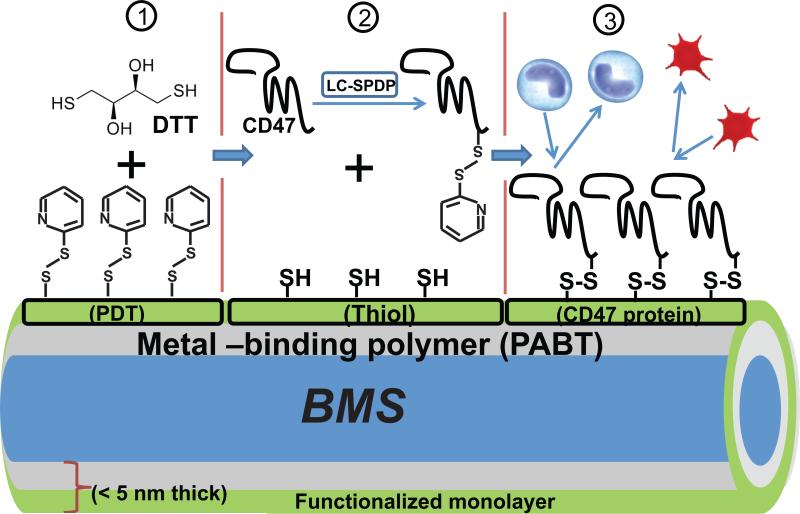
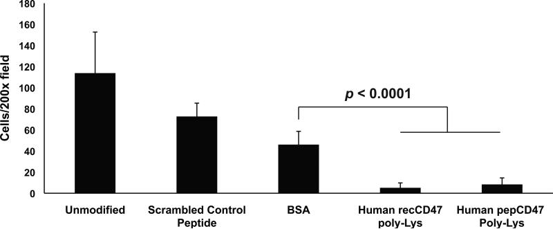
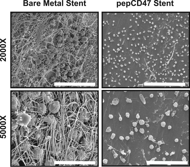
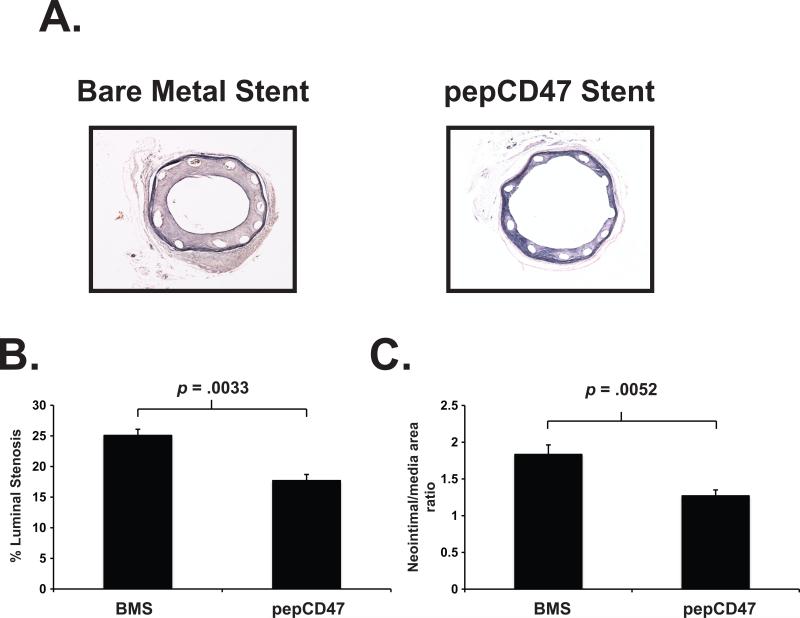
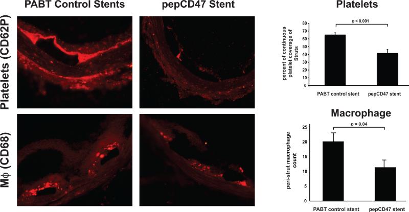
The effectiveness of endovascular stents is hindered by in-stent restenosis (ISR), a secondary re-obstruction of treated arteries due to unresolved inflammation and activation of smooth muscle cells in the arterial wall. We previously demonstrated that immobilized CD47, a ubiquitously expressed transmembrane protein with an established role in immune evasion, can confer biocompatibility when appended to polymeric surfaces. In present studies, we test the hypothesis that CD47 immobilized onto metallic surfaces of stents can effectively inhibit the inflammatory response thus mitigating ISR. Recombinant CD47 (recCD47) or a peptide sequence corresponding to the Ig domain of CD47 (pepCD47), were attached to the surfaces of both 316L-grade stainless steel foils and stents using bisphosphonate coordination chemistry and thiol-based conjugation reactions to assess the anti-inflammatory properties of CD47-functionalized surfaces. Initial in vitro and ex vivo analysis demonstrated that both recCD47 and pepCD47 significantly reduced inflammatory cell attachment to steel surfaces without impeding on endothelial cell retention and expansion. Using a rat carotid stent model, we showed that pepCD47-functionalized stents prevented fibrin and platelet thrombus deposition, inhibited inflammatory cell attachment, and reduced restenosis by 30%. It is concluded that CD47-modified stent surfaces mitigate platelet and inflammatory cell attachment, thereby disrupting ISR pathophysiology.

Keywords: Bare metal stents; Bioactive surface coating; Inflammation; Platelets; Restenosis; Signal regulatory protein alpha.


Figures

Fig. 1
Fig. 1
Schematic representation of modification chemistry used to append CD47 to bare metal stents (BMS). After modification of steel surfaces with polallylamine bisphosphonate comprising latent thiol groups, PABT and deprotection of latent thiols, the surface was rendered thiol-reactive due to installment of pyridyldithio (PDT) groups using PEI-PDT (step 1). Since only a minor fraction of PDT groups in PEI-PDT is consumed in the reaction with thiols, PEI-PDT acts as an amplifier of thiol groups which are formed during reduction of PDT groups with dithiothreitol, DTT (step 2). In parallel, recCD47 or pepCD47 is modified at poly(Lys) segment with bifunctional (amine-, thiol-reactive) cross-linker, Sulfo-LC-SPDP (step 2). Finally, the thiol-reactive protein moiety is attached via disulfide bridges to the functionalized surface in the course of reaction between surface thiols and protein-bound PDT groups (step 3).
Fig. 2
Fig. 2
CD47 Quantification. (A) The modification chemistry summarized in Figure 1 was utilized to facilitate quantification. Following PEI-PDT addition, primary amines were acetylated using sulfo-NHS-Acetate. recCD47 and pepCD47 were biotinylated using Sulfo-NHS-LC-LC-Biotin. Biotinylated recCD47 and pepCD47 were then reacted with the steel surfaces. ABC reagent and Ultra TMB substrate were used to determine the concentration of CD47 by comparison to a standard curve (inset) prepared using escalating amount of biotinylated IgG assayed in the same manner. (B) CD47 concentration on steel surfaces in the presence or absence of bi-functional cross-linking agent (sulfo-LC-SPDP).
Fig. 3
Fig. 3
CD47 immobilization prevents attachment of cultured monocyte-derived macrophages to steel surfaces. THP-1 cells were differentiated with the addition of PMA to their media and were cultured on steel foils for 3 days. After 3 days, the foils were gently rinsed with DPBS, fixed with 4% formaldehyde, and nuclei stained with DAPI. DAPI staining of adherent cells was visualized and randomly selected 200X fields were counted. Results represent the average and standard deviation of the mean of nine individual fields (P < 0.0001).
Fig. 4
Fig. 4
CD47 immobilization prevents the attachment of blood leukocytes to stainless steel surfaces. The Chandler Loop apparatus was used, as detailed in Materials and Methods, to expose citrate-treated whole human blood to the luminal surface of unmodified steel inserts or BSA, recCD47, or pepCD47-modified steel inserts that were placed into medical grade PVC tube conduits. DAPI staining of adherent cells were visualized, and randomly selected 200X fields were counted. Results show that immobilization of CD47 significantly reduced blood cell adhesion to the steel surfaces. Results represent the average and standard deviation of the mean of nine individual fields (P < 0.0001).
Fig. 5
Fig. 5
Comparative analysis of the effect of rat CD47 immobilization strategy upon blood cell attachment. Thiolated or poly-lysine modified pepCD47, derived from the rat CD47 sequence, were appended to steel inserts as detailed in Materials and Methods. Heparinized whole rat blood was perfused, via the Chandler Loop Apparatus, over the luminal surface of unmodified steel inserts or BSA, pepCD47-modified, or recCD47-modified steel inserts, which were placed into medical grade PVC tube conduits. After three hours, the inserts were removed and processed as detailed in Materials and Methods. Following DAPI staining, nine randomly selected fields of view under 200X magnification were counted and statistical analyses were performed. Results represent the average and standard deviation of the mean of nine individual fields (P < 0.0001).
Fig. 6
Fig. 6
Endothelial cell retention, under physiologically relevant shear flow, is not affected by CD47 immobilization. (A) Representative fluorescent micrographs of HUVECs cultured on thiolated pepCD47, recCD47, BSA-modified and unmodified stainless steel surfaces. Once confluent, the HUVECs were exposed to 25 dynes/cm2 fluid shear stress (FSS) for 4 hr using the FlexCell® Streamer® device. Following shear stress exposure, the HUVECs were fixed and permeabilized. Actin stress fibers were then stained with rhodamine-Phalloidin, following which the nuclei were DAPI stained. (B) DAPI positive cells from nine random fields were counted and compared with cell counts from cultured HUVECs grown on the various surface, but not exposed to FSS. Graph shows percent retention for each surface tested.
Fig. 7
Fig. 7
Surface characterization of explanted endovascular stent following 30-minute deployment in a rat carotid artery. Representative scanning electron micrographs of the surface of bare metal stents or stents coated with thiolated pepCD47. Images show large fibrin deposition and trapped cells on the unmodified stents surface. In contrast pepCD47 stents exhibited less fibrin on the surface and the attached cells appeared to be more rounded and less spread. Scale Bar = 30 μm (2000X) and 10 μm (5000X)
Fig. 8
Fig. 8
The anti-restenotic effect of CD47 modified stents in a rat carotid artery. Bare metal stents or thiolated pepCD47-modified stents were deployed in the carotid artery of rats. The stents were explanted after 14 days. (A) Representative Hematoxylin-eosin stain of explanted stents. Quantitative morphometric analyses of explanted stents demonstrate that (B) luminal stenosis and (C) Neointima/media area ratio are significantly (p ≤ 0.05) reduced as a function of CD47 immobilization. Data represent measurements from 10 individual explanted stents and are presented as mean ± SD.
Fig. 9
Fig. 9
Temporal qualitative and quantitative comparisons of cell types from explanted endovascular stents. Representative fluorescent micrographs of explanted bare metal stents, modified with the chemical crosslinker (PABT) or with thiolated pepCD47, stained for surface markers of platelets (CD62P) or macrophages (CD68) after 30 minutes or 3 days respectively showing cellular interactions at the stent strut(s). Scale bar = 100μm. Data represent the mean from 2-3 struts from explanted stents and are presented as mean ± SD.

References

    1. Mozaffarian D, Benjamin EJ, Go AS, Arnett DK, Blaha MJ, Cushman M, et al. Heart disease and stroke statistics--2015 update: a report from the American Heart Association. Circulation. 2015;131:e29–322. - PubMed
    1. Butt M, Connolly D, Lip GY. Drug-eluting stents: a comprehensive appraisal. Future cardiology. 2009;5:141–57. - PubMed
    1. Simard T, Hibbert B, Ramirez FD, Froeschl M, Chen YX, O'Brien ER. The evolution of coronary stents: a brief review. Can J Cardiol. 2014;30:35–45. - PubMed
    1. Rathore S, Terashima M, Katoh O, Matsuo H, Tanaka N, Kinoshita Y, et al. Predictors of angiographic restenosis after drug eluting stents in the coronary arteries: contemporary practice in real world patients. EuroIntervention. 2009;5:349–54. - PubMed
    1. Jaffe R, Strauss BH. Late and very late thrombosis of drug-eluting stents: evolving concepts and perspectives. J Am Coll Cardiol. 2007;50:119–27. - PubMed

Publication types

MeSH terms