Comparative Study
. 2016 Apr 15;122(8):1216-27.
doi: 10.1002/cncr.29912. Epub 2016 Feb 23.

Metformin prevents hepatocellular carcinoma development by suppressing hepatic progenitor cell activation in a rat model of cirrhosis

Affiliations
Comparative Study

Metformin prevents hepatocellular carcinoma development by suppressing hepatic progenitor cell activation in a rat model of cirrhosis

Danielle K DePeralta et al. Cancer. .

Abstract

Background: Hepatocellular carcinoma (HCC)-associated mortality is increasing at an alarming rate, and there is a readily identifiable cohort of at-risk patients with cirrhosis, viral hepatitis, nonalcoholic fatty liver disease, and diabetes. These patients are candidates for chemoprevention. Metformin is an attractive agent for chemoprevention because it is inexpensive, has a favorable safety profile, and is well tolerated over long time periods.

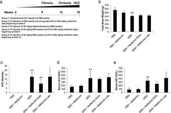
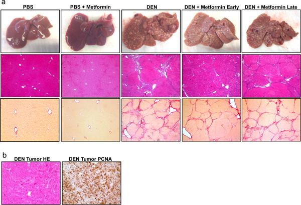
Methods: The authors studied the efficacy of metformin as a prevention agent in a clinically relevant rat model of HCC, in which tumors develop in the setting of chronic inflammation and cirrhosis. Repeated injections of diethylnitrosamine were used to induce sequential cirrhosis and HCC, and metformin was administered at the first signs of either fibrosis or cirrhosis.

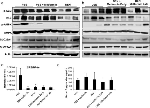
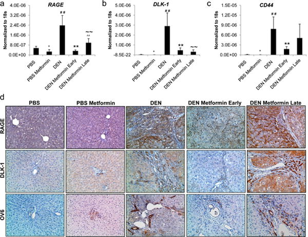
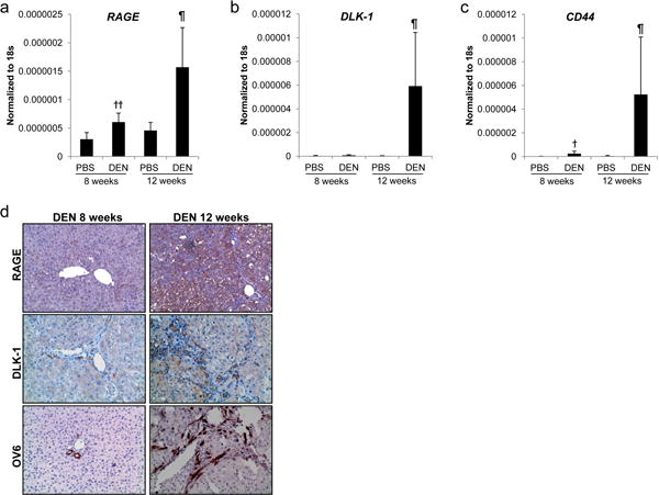
Results: Prolonged metformin exposure was safe and was associated with decreases in fibrotic and inflammatory markers, especially when administered early at the first signs of fibrosis. In addition, early metformin treatment led to a 44% decrease in HCC incidence, whereas tumor burden was unchanged when metformin was administered at the first signs of cirrhosis. It is noteworthy that activation of the hepatic progenitor/stem cell compartment was first observed at the onset of cirrhosis; therefore, only early metformin treatment suppressed receptor for advanced glycation end products and inhibited the activation of hepatic progenitor cells.

Conclusions: The current results are the first to demonstrate an effect on progenitor/stem cells in the setting of chemoprevention and provide further rationale to explore metformin as an early intervention in clinical trials of patients with chronic liver disease at high risk for HCC.

Keywords: hepatocellular carcinoma (HCC); liver; oval cells; prevention; receptor for advanced glycation end products (RAGE).


Conflict of interest statement

Conflicts: The authors declare no conflict of interest.

Figures

Figure 1
Figure 1. Early metformin treatment inhibits hepatocellular tumorigenesis in a cirrhosis model
(a) Rats were treated with weekly i.p. diethylnitrosamine (DEN) in order to induce sequential fibrosis, cirrhosis, and HCC. Control rats received saline (PBS). PBS control rats received daily treatment with metformin by oral gavage after 8 weeks (PBS + metformin) while DEN-injured rats received metformin treatment after either 8 weeks (DEN + Metformin Early) or 12 weeks (DEN + Metformin Late) coinciding with the onset of fibrosis or cirrhosis, respectively. Rats were sacrificed at 18 weeks (n = 9 for each group). (b) Total body weight and (c) the number of surface tumors were measured for each animal at the time of sacrifice. Serum levels of (d) alkaline phosphatase (ALP) and (e) aspartate transaminase (AST) were measured to assess hepatocellular injury (n = 5 for each group). + p < 0.05 PBS compared to PBS Metformin, ## p < 0.01 PBS compared to DEN, ** p < 0.01 DEN compared to DEN + Metformin Early, and ˆ p < 0.05 DEN + Metformin Early compared to DEN + Metformin Late.
Figure 2
Figure 2. Early metformin treatment improves liver disease in DEN-injured rats
(a) Representative macroscopic pictures of livers (top panel), H&E staining (40×, middle panel) and Sirius red staining (40×, bottom panel) from PBS, PBS + Metformin, DEN, DEN + Metformin Early, and DEN + Metformin Late. (b) H&E staining (100X, left picture) and PCNA staining (100×, right picture) of a HCC from a DEN-injured liver.
Figure 3
Figure 3. Early metformin treatment decreases liver fibrosis in DEN-injured rats
(a) The collagen proportional area (CPA) was quantified for every animal in each group with ImageJ analysis software. (b) The Ishak score for each animal was determined by a blinded, board-certified pathologist. Serum levels of (c) albumin (Alb) and (d) total bilirubin (TBIL) were measured to assess liver function (n = 5 for each group). (e) The extent of inflammation for each animal was determined by a blinded, board-certified pathologist. ## p < 0.01 PBS compared to DEN, * p < 0.05 and ** p < 0.01 DEN compared to DEN + Metformin Early, and ˆ p < 0.05 and ˆˆ p < 0.01 DEN + Metformin Early compared to DEN + Metformin Late.
Figure 4
Figure 4. Metformin does not inhibit hepatic lipogenesis
Representative western blot analysis of surrounding liver tissue from (a) PBS, PBS + Metformin, and DEN animals and (b) DEN, DEN + Metformin Early, and DEN + Metformin Late animals. Western blots were probed for p-ACC, total ACC, p-AMPK, total AMPK, SLC22A1, and SLC22A3. β-actin was used as a loading control. Western blots were repeated at least 3 times to ensure reproducibility. (c) The mRNA expression of SREBP-1c in surrounding liver tissue was assessed by real-time PCR using Taqman primers (n = 5 for each group). (d) Serum levels of triglycerides were measured (n = 5 for each group). ++ p < 0.01 PBS compared to PBS Metformin, and # p < 0.05 PBS compared to DEN.
Figure 5
Figure 5. Early metformin treatment decreases hepatic progenitor cell activation
The mRNA expression of (a) RAGE, (b) DLK-1 and (c) CD44 in surrounding liver tissue from PBS, PBS + Metformin, DEN, DEN + Metformin Early, and DEN + Metformin Late animals was assessed by real-time PCR using Taqman primers (n = 5 for each group). (d) Immunohistochemistry was performed for RAGE (top panel), DLK-1 (middle panel) and OV-6 (bottom panel) in surrounding liver tissue from the same groups. + p < 0.05 PBS compared to PBS Metformin, ## p < 0.01 PBS compared to DEN, ** p < 0.01 DEN compared to DEN + Metformin Early, ~~ p < 0.01 DEN compared to DEN + Metformin Late, and ˆˆ p < 0.01 DEN + Metformin Early compared to DEN + Metformin Late.
Figure 6
Figure 6. Hepatic progenitor cells activate at the onset of cirrhosis
The mRNA expression of (a) RAGE, (b) DLK-1 and (c) CD44 in surrounding liver tissue from PBS control and DEN-injured animals after 8 and 12 weeks of treatment was assessed by real-time PCR using Taqman primers (n = 5 for each group). (d) Immunohistochemistry was performed for RAGE (top panel), DLK-1 (middle panel) and OV-6 (bottom panel) in surrounding liver tissue from DEN-injured animals after 8 and 12 weeks. † p < 0.05 and †† p < 0.01 PBS 8 weeks compared to DEN 8 weeks, and ¶ p < 0.05 DEN 12 weeks compared to all groups.

References

    1. Jemal A, Bray F, Center MM, Ferlay J, Ward E, Forman D. Global cancer statistics. CA Cancer J Clin. 2011;61:69–90. - PubMed
    1. El-Serag HB. Hepatocellular carcinoma. N Engl J Med. 2011;365:1118–1127. - PubMed
    1. Polesel J, Zucchetto A, Montella M, et al. The impact of obesity and diabetes mellitus on the risk of hepatocellular carcinoma. Ann Oncol. 2009;20:353–357. - PubMed
    1. Evans JM, Donnelly LA, Emslie-Smith AM, Alessi DR, Morris AD. Metformin and reduced risk of cancer in diabetic patients. BMJ. 2005;330:1304–1305. - PMC - PubMed
    1. Decensi A, Puntoni M, Goodwin P, et al. Metformin and cancer risk in diabetic patients: a systematic review and meta-analysis. Cancer Prev Res. 2010;3:1451–1461. - PubMed

Publication types

MeSH terms