

Strength of the porcine proximal femoral epiphyseal plate: the effect of different loading directions and the role of the perichondrial fibrocartilaginous complex and epiphyseal tubercle - an experimental biomechanical study
- PMID: 26914749
- PMCID: PMC4648830
- DOI: 10.1186/s40634-014-0004-y
Strength of the porcine proximal femoral epiphyseal plate: the effect of different loading directions and the role of the perichondrial fibrocartilaginous complex and epiphyseal tubercle - an experimental biomechanical study
Abstract
Background: The high loads on adolescent athletes' musculoskeletal system are known to cause morphological and degenerative changes in bone, intervertebral discs and joints. It has been suggested that the cam deformity of the proximal femoral head originates from a subclinical slipped capital femoral epiphysis (SCFE) as a result of non-physiological loading. The perichondrial fibrocartilaginous complex (PFC) and the epiphyseal tubercle are believed to stabilise the proximal femoral epiphysis, but their role is still unclear. The aim of the present study was to develop an experimental, biomechanical model to evaluate the strength of the porcine proximal femoral epiphysis in different loading directions and, furthermore, to investigate the stabilising role of the PFC and the epiphyseal tubercle.
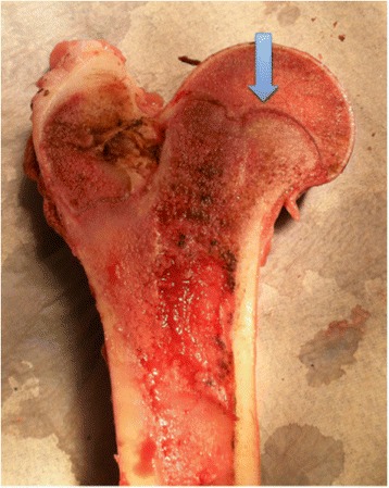
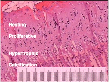
Methods: A descriptive laboratory study. An in-vitro model was developed and nine young (5 months) porcine proximal femoral epiphyses were loaded to failure; three in the anterior-posterior direction, three in the lateral-medial direction and three in the vertical direction. The injured proximal femoral epiphyses were then examined both macroscopically and histologically.
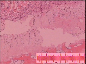
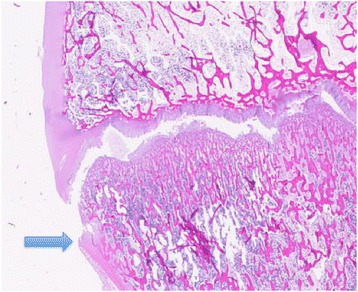
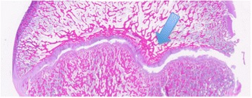
Results: Anterior and lateral loading of the proximal femoral epiphysis resulted in failure of the epiphyseal plate, while vertical loading resulted in a fracture epiphyseolysis. The epiphysis was weakest when exposed to a lateral load and strongest when exposed to a vertical load. Despite histological epiphyseolysis, the PFC was intact in 15 of 27 (56%) slices. In histological examinations, the epiphyseal tubercle appears to halt the slide of the epiphysis.
Conclusions: We have developed an experimental, biomechanical model to measure the strength of the proximal femoral epiphyseal plate in different loading directions. The strength of the proximal femur was weakest through the epiphyseal plate. The epiphysis was weakest when exposed to a lateral load and strongest when exposed to a vertical load. The epiphyseal tubercle and the PFC stabilise the epiphysis when the epiphyseal plate is damaged. The findings in the present study indicate that overloading the hips in growing individuals can disrupt the epiphyseal plate. These findings may have implications when it comes to understanding the pathogenesis of cam deformity of the hip.
Keywords: Epiphyseal plate; Epiphyseal tubercle; Epiphysiolysis; Hip; Load; Perichondrial fibrocartilaginous complex; Porcine.
Figures


Similar articles
-
Effects of joint loading on the development of capital femoral epiphysis morphology.Arch Orthop Trauma Surg. 2023 Sep;143(9):5457-5466. doi: 10.1007/s00402-023-04795-0. Epub 2023 Mar 1. Arch Orthop Trauma Surg. 2023. PMID: 36856839
-
Smaller Epiphyseal Tubercle and Larger Peripheral Cupping in Slipped Capital Femoral Epiphysis Compared with Healthy Hips: A 3-Dimensional Computed Tomography Study.J Bone Joint Surg Am. 2020 Jan 2;102(1):29-36. doi: 10.2106/JBJS.19.00291. J Bone Joint Surg Am. 2020. PMID: 31596801
-
Morphologic Features of the Contralateral Femur in Patients With Unilateral Slipped Capital Femoral Epiphysis Resembles Mild Slip Deformity: A Matched Cohort Study.Clin Orthop Relat Res. 2018 Apr;476(4):890-899. doi: 10.1007/s11999.0000000000000127. Clin Orthop Relat Res. 2018. PMID: 29481345 Free PMC article.
-
Increased body mass index percentile is associated with decreased epiphyseal tubercle size in asymptomatic children and adolescents with healthy hips.J Child Orthop. 2020 Jun 1;14(3):167-174. doi: 10.1302/1863-2548.14.200042. J Child Orthop. 2020. PMID: 32582383 Free PMC article. Review.
-
[Slipped capital femoral epiphysis-etiology and pathogenesis].Orthopade. 2019 Aug;48(8):644-650. doi: 10.1007/s00132-019-03743-4. Orthopade. 2019. PMID: 31115600 Review. German.
Cited by
-
The metaphyseal fossa surrounding the epiphyseal tubercle is larger in hips with moderate and severe slipped capital femoral epiphysis than normal hips.J Child Orthop. 2020 Jun 1;14(3):184-189. doi: 10.1302/1863-2548.14.200010. J Child Orthop. 2020. PMID: 32582385 Free PMC article. Review.
-
Age- and Sex-Specific Morphologic Variations of Capital Femoral Epiphysis Growth in Children and Adolescents Without Hip Disorders.Orthop J Sports Med. 2018 Jun 25;6(6):2325967118781579. doi: 10.1177/2325967118781579. eCollection 2018 Jun. Orthop J Sports Med. 2018. PMID: 30090833 Free PMC article.
-
Effects of joint loading on the development of capital femoral epiphysis morphology.Arch Orthop Trauma Surg. 2023 Sep;143(9):5457-5466. doi: 10.1007/s00402-023-04795-0. Epub 2023 Mar 1. Arch Orthop Trauma Surg. 2023. PMID: 36856839
-
Stressed or fractured: MRI differentiating indicators of physeal injury.Skeletal Radiol. 2024 Nov;53(11):2437-2447. doi: 10.1007/s00256-024-04670-y. Epub 2024 Apr 1. Skeletal Radiol. 2024. PMID: 38557698
-
The surgical destabilization of the abductor muscle leads to development of instability-associated hip osteoarthritis in mice.J Hip Preserv Surg. 2023 Jul 29;10(3-4):158-165. doi: 10.1093/jhps/hnad015. eCollection 2023 Aug-Dec. J Hip Preserv Surg. 2023. PMID: 38162262 Free PMC article.
References
-
- Arkin AM, Katz JF. The effects of pressure on epiphyseal growth; the mechanism of plasticity of growing bone. J Bone Joint Surg Am. 1956;38-A(5):1056–1076. - PubMed
-
- Malina RM. Exercise as an influence upon growth. Review and critique of current concepts. Clinical pediatrics. 1969;8(1):16–26. - PubMed
-
- Strobino LJ, French GO, Colonna PC. The effect of increasing tensions on the growth of epiphyseal bone. Surgery, gynecology & obstetrics. 1952;95(6):694–700. - PubMed
LinkOut - more resources
Full Text Sources
Other Literature Sources
Miscellaneous
