

Cyclical loading causes injury in and around the porcine proximal femoral physeal plate: proposed cause of the development of cam deformity in young athletes
- PMID: 26914874
- PMCID: PMC4545757
- DOI: 10.1186/s40634-015-0022-4
Cyclical loading causes injury in and around the porcine proximal femoral physeal plate: proposed cause of the development of cam deformity in young athletes
Abstract
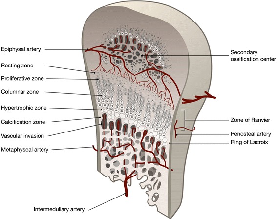
Background: The repetitive load to which the adolescent athlete's body is exposed during training and competition affects bone growth. In previous studies, abnormalities of the spine and extremities of adolescent athletes have been described on radiographs and this also applies to the hip. The cam deformity of the hip is an extension of the physeal plate and develops during the adolescent athlete's growth. Studies of the porcine spine have shown that the vertebral endplates, apophyseal rings and intervertebral discs are susceptible to both static and repetitive loads. The proximal physeal plate of the porcine femur is susceptible to static loads, but no studies have been performed on its susceptibility to repetitive loads. The purpose of this study was to investigate the susceptibility of the proximal porcine femur to repetitive loads.
Methods: Descriptive laboratory study. Seven proximal femurs from four young (5 months) pigs were loaded repetitively (50,000 cycles) using a previously developed model. Three were loaded vertically, three antero-superiorly and one was used as a control. All femurs were examined macroscopically, histologically and with MRI after loading.
Results: No macroscopic injuries were detected on any of the femurs after loading. Fluid redistribution was seen in all femurs on MRI compared with the unloaded control. Injuries were seen in all loaded femurs on microscopic examination of histological samples. Injuries, perpendicularly to the physeal plate and fractures adjacent to the plate, were seen in the vertically loaded specimens. In the antero-superiorly loaded specimen, the injury in the growth plate was parallel to the plate.
Conclusion: Repeated loading of the young porcine hip leads to histological injuries in and adjacent to the physeal plate. These injuries are likely to cause growth disturbances in the proximal femur. We propose that such injuries may be induced in adolescent athletes and offer a plausible explanation for the development of the cam deformity.
Keywords: Adolescent; Athlete; Biomechanics; Cam; Femoroacetabular impingement; Hip; Physeal plate; Porcine.
Figures


References
-
- Agricola R, Heijboer MP, Ginai AZ, Roels P, Zadpoor AA, Verhaar JA, Weinans H, Waarsing JH. A cam deformity is gradually acquired during skeletal maturation in adolescent and young male soccer players: a prospective study with minimum 2-year follow-up. Am J Sports Med. 2014;42(4):798–806. doi: 10.1177/0363546514524364. - DOI - PubMed
-
- Baranto A, Ekstrom L, Hellstrom M, Lundin O, Holm S, Sward L. Fracture patterns of the adolescent porcine spine: an experimental loading study in bending-compression. Spine (Phila Pa 1976) 2005;30(1):75–82. - PubMed
-
- Baranto A, Ekstrom L, Holm S, Hellstrom M, Hansson HA, Sward L. Vertebral fractures and separations of endplates after traumatic loading of adolescent porcine spines with experimentally-induced disc degeneration. Clin Biomech (Bristol, Avon) 2005;20(10):1046–1054. doi: 10.1016/j.clinbiomech.2005.06.014. - DOI - PubMed
LinkOut - more resources
Full Text Sources
Other Literature Sources
