

Overexpression of microRNA-99a Attenuates Cardiac Hypertrophy
- PMID: 26914935
- PMCID: PMC4767297
- DOI: 10.1371/journal.pone.0148480
Overexpression of microRNA-99a Attenuates Cardiac Hypertrophy
Abstract
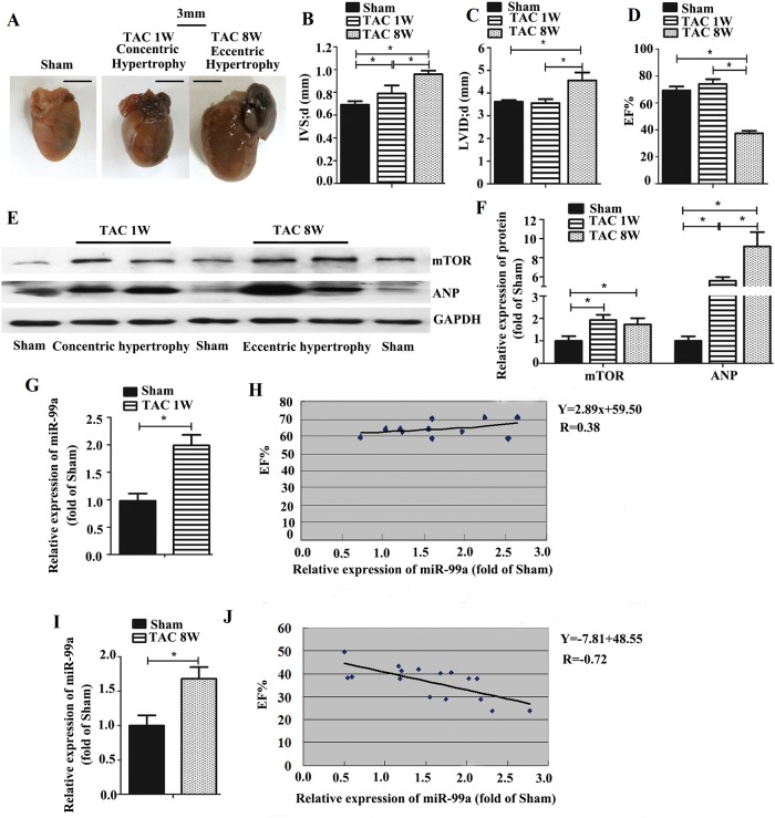
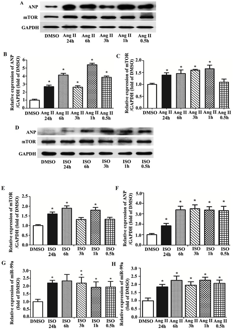
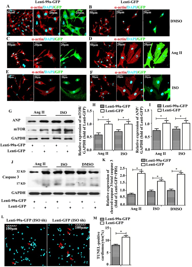
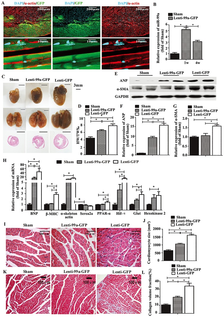
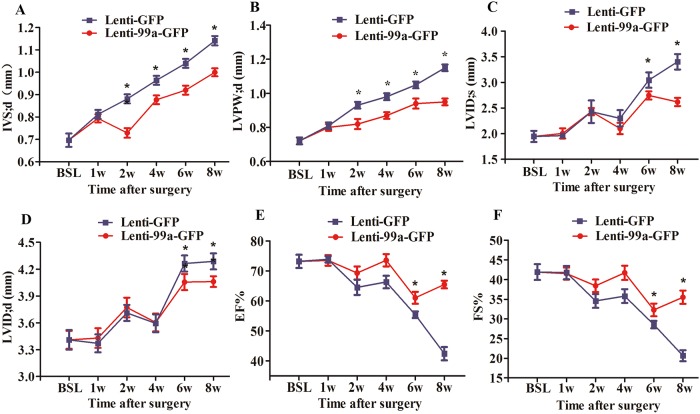
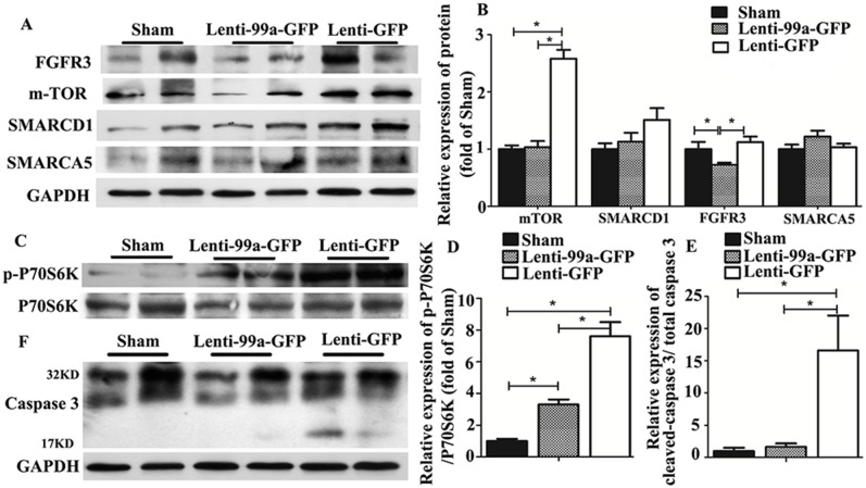
Pathological cardiomyocyte hypertrophy is associated with significantly increased risk of heart failure, one of the leading medical causes of mortality worldwide. MicroRNAs are known to be involved in pathological cardiac remodeling. However, whether miR-99a participates in the signaling cascade leading to cardiac hypertrophy is unknown. To evaluate the role of miR-99a in cardiac hypertrophy, we assessed the expression of miR-99a in hypertrophic cardiomyocytes induced by isoprenaline (ISO)/angiotensin-II (Ang II) and in mice model of cardiac hypertrophy induced by transverse aortic constriction (TAC). Expression of miR-99a was evaluated in these hypertrophic cells and hearts. We also found that miR-99a expression was highly correlated with cardiac function of mice with heart failure (8 weeks after TAC surgery). Overexpression of miR-99a attenuated cardiac hypertrophy in TAC mice and cellular hypertrophy in stimuli treated cardiomyocytes through down-regulation of expression of mammalian target of rapamycin (mTOR). These results indicate that miR-99a negatively regulates physiological hypertrophy through mTOR signaling pathway, which may provide a new therapeutic approach for pressure-overload heart failure.
Conflict of interest statement
Figures
Similar articles
-
MicroRNA-92b-3p suppresses angiotensin II-induced cardiomyocyte hypertrophy via targeting HAND2.Life Sci. 2019 Sep 1;232:116635. doi: 10.1016/j.lfs.2019.116635. Epub 2019 Jul 5. Life Sci. 2019. PMID: 31283925
-
MicroRNA-17-5p Promotes Cardiac Hypertrophy by Targeting Mfn2 to Inhibit Autophagy.Cardiovasc Toxicol. 2021 Sep;21(9):759-771. doi: 10.1007/s12012-021-09667-w. Epub 2021 Jun 12. Cardiovasc Toxicol. 2021. PMID: 34120306
-
Myocyte-specific enhancer factor 2C: a novel target gene of miR-214-3p in suppressing angiotensin II-induced cardiomyocyte hypertrophy.Sci Rep. 2016 Oct 31;6:36146. doi: 10.1038/srep36146. Sci Rep. 2016. PMID: 27796324 Free PMC article.
-
The function of miRNA in cardiac hypertrophy.Cell Mol Life Sci. 2012 Nov;69(21):3561-70. doi: 10.1007/s00018-012-1126-y. Epub 2012 Aug 25. Cell Mol Life Sci. 2012. PMID: 22926414 Free PMC article. Review.
-
MicroRNAs in Cardiac Hypertrophy.Int J Mol Sci. 2019 Sep 23;20(19):4714. doi: 10.3390/ijms20194714. Int J Mol Sci. 2019. PMID: 31547607 Free PMC article. Review.
Cited by
-
The Impact of microRNAs in Renin-Angiotensin-System-Induced Cardiac Remodelling.Int J Mol Sci. 2021 Apr 30;22(9):4762. doi: 10.3390/ijms22094762. Int J Mol Sci. 2021. PMID: 33946230 Free PMC article. Review.
-
Mybl2 rejuvenates heart explant-derived cells from aged donors after myocardial infarction.Aging Cell. 2020 Jul;19(7):e13174. doi: 10.1111/acel.13174. Epub 2020 Jun 19. Aging Cell. 2020. PMID: 32558221 Free PMC article.
-
Circulating microRNAs and therapy-associated cardiac events in HER2-positive breast cancer patients: an exploratory analysis from NeoALTTO.Breast Cancer Res Treat. 2024 Jul;206(2):285-294. doi: 10.1007/s10549-024-07299-6. Epub 2024 Apr 30. Breast Cancer Res Treat. 2024. PMID: 38689174 Free PMC article.
-
Developmental programming: Adipose depot-specific regulation of non-coding RNAs and their relation to coding RNA expression in prenatal testosterone and prenatal bisphenol-A -treated female sheep.Mol Cell Endocrinol. 2023 Mar 15;564:111868. doi: 10.1016/j.mce.2023.111868. Epub 2023 Jan 26. Mol Cell Endocrinol. 2023. PMID: 36708980 Free PMC article.
-
Nogo-C regulates cardiomyocyte apoptosis during mouse myocardial infarction.Cell Death Dis. 2016 Oct 20;7(10):e2432. doi: 10.1038/cddis.2016.331. Cell Death Dis. 2016. PMID: 27763637 Free PMC article.
References
-
- Bonnin CM, Sparrow MP, Taylor RR. Increased protein synthesis and degradation in the dog heart during thyroxine administration. (1983) J Mol Cell Cardiol 15: 245–250. - PubMed
-
- Morgan HE, Gordon EE, Kira Y, Chua HL, Russo LA, Peterson CJ, et al. (1987) Biochemical mechanisms of cardiac hypertrophy. Annu Rev Physiol 49: 533–543. - PubMed
-
- Dorn GW 2nd, Robbins J, Sugden PH. (2003) Phenotyping hypertrophy: eschew obfuscation. Circ Res. 92: 1171–1175. - PubMed
Publication types
MeSH terms
Substances
LinkOut - more resources
Full Text Sources
Other Literature Sources
Miscellaneous
