Review
. 2016 Apr;18(4):33.
doi: 10.1007/s11886-016-0712-2.

Impact of Right-Sided-Catheter-Based Valve Implantation on Decision-Making in Congenital Heart Disease

Affiliations
Review

Impact of Right-Sided-Catheter-Based Valve Implantation on Decision-Making in Congenital Heart Disease

Joanna Ghobrial et al. Curr Cardiol Rep. 2016 Apr.

Abstract

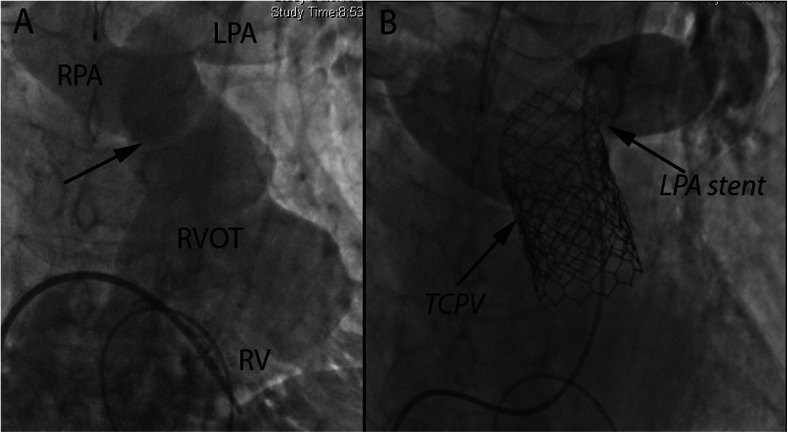
There is a growing appreciation for the adverse long-term impact of right-sided valvular dysfunction in patients with congenital heart disease. Although right-sided valvular stenosis and/or regurgitation is often better tolerated than left-sided valvular dysfunction in the short and intermediate term, the long-term consequences are numerous and include, but are not limited to, arrhythmias, heart failure, and multi-organ dysfunction. Surgical right-sided valve interventions have been performed for many decades, but the comorbidities associated with multiple surgeries are a concern. Transcatheter right-sided valve replacement is safe and effective and is being performed at an increasing number of centers around the world. It offers an alternative to traditional surgical techniques and may potentially alter the decision making process whereby valvular replacement is performed prior to the development of long-term sequelae of right-sided valvular dysfunction.

Keywords: Congenital heart disease; Melody valve; Pulmonary regurgitation; Pulmonary stenosis; Sapien valve; Tetralogy of Fallot; Transcatheter valve replacement; Tricuspid regurgitation; Tricuspid stenosis.


Figures

Fig. 1
Fig. 1
a Transthoracic echocardiographic image (parasternal short axis view) of the right ventricular outflow tract (RVOT) showing turbulent color Doppler flow across a stenotic pulmonary valve. AV = aortic valve. b Parasternal short axis view of RVOT with an indwelling stent in the main pulmonary artery well above the level of the pulmonary valve annulus and color Doppler flow showing severe pulmonary valve regurgitation (PR). Note the regurgitant reverse color Doppler flow in the left pulmonary artery (LPA) and right pulmonary artery (RPA). c Parasternal short axis view of the RVOT showing a Melody valve successfully deployed in the MPA
Fig. 2
Fig. 2
a Antero-posterior (AP) angiographic view of a bioprosthetic pulmonary valve (BPV) with contrast injection in the pulmonary artery showing severe pulmonary regurgitation and a stent in the right pulmonary artery. b AP view of the same bioprosthetic valve after transcatheter pulmonary valve placement (TPV) of a Melody valve. No residual pulmonary regurgitation is seen
Fig. 3
Fig. 3
a Antero-posterior and cranial projection of a contrast injection into the main pulmonary artery (MPA) demonstrating severe pulmonary regurgitation into the native right ventricular outflow tract (RVOT). The right and left pulmonary arteries (RPA and LPA) are labeled, with a narrowing just proximal to the branching of the MPA measuring approximately 14.9 mm. Note sternal wires from previous median sternotomy. b Bare metal stent placed across the narrowed MPA. c Image following Melody transcatheter pulmonary valve (TPV) placement within the stent. Contrast injection in the MPA shows no residual pulmonary regurgitation
Fig. 4
Fig. 4
a Lateral angiographic view of a native right ventricular outflow tract (RVOT) with a wire placed in the distal left pulmonary artery and catheter in a dilated main pulmonary artery (MPA) showing severe regurgitation across a dysfunctional native pulmonary valve (PV). b Antero-posterior (AP) angiographic view of the same native RVOT showing severe PR. c AP fluoroscopic view of the Sapien XT transcatheter pulmonary valve (TPV) positioned within a stent across the native pulmonary valve. d Lateral view of the Sapien valve delivery system positioned across the pre-stented PV. e AP view of deployed Sapien valve within the stented PV. f Lateral view of the deployed Sapien valve within the pre-stented PV
Fig. 5
Fig. 5
a Antero-posterior (AP) view of a fractured Melody transcatheter pulmonary valve (TPV) within a right ventricular to pulmonary artery homograft. b AP view of a deployed Melody TPV within the previously fractured Melody valve after pre-stenting. c Transthoracic echocardiographic image with continuous wave (CW) Doppler across the fractured Melody TPV demonstrating moderate stenosis (peak velocity 3.6 m/s, peak gradient of 53 mmHg, and mean gradient 29 mmHg). d Transthoracic echocardiographic image with CW Doppler signal across the deployed Melody valve within the previously fractured Melody valve showing significant improvement (reduction in velocity to 2.3 m/s, peak gradient to 22 mmHg and mean gradient to 14 mmHg)
Fig. 6
Fig. 6
Antero-posterior (AP) angiographic view of aortic root injection with simultaneous balloon inflation across the RVOT showing coronary artery compression (black arrow) of an anomalous left anterior descending artery
Fig. 7
Fig. 7
a Antero-posterior (AP) angiographic view of aortic root injection prior to balloon inflation within a dysfunctional bioprosthetic pulmonary valve. b Aortic root injection with simultaneous balloon inflation across the pulmonary valve showing aortic root compression and the development of aortic regurgitation (black arrow)
Fig. 8
Fig. 8
a Antero-posterior (AP) and cranial (CRA) angiographic view of a patient with transannular patch repair of the right ventricular outflow tract (RVOT) and main pulmonary artery (MPA) with a relative narrowing (black arrow) proximal to the bifurcation. The right ventricle (RV) is severely dilated. Right pulmonary arteries = RPA; left pulmonary artery = LPA. b Following pre-stenting with two stents, one extending into the LPA and “jailing” the RPA, but with open stent struts allowing antegrade bloodflow into the RPA. The Sapien transcatheter pulmonary valve (TPV) placement was deployed within the two stents. This technique allows for anchoring of the TPV in dilated RVOTs. Contrast injection in the LPA demonstrates a competent TPV without significant pulmonary regurgitation
Fig. 9
Fig. 9
a Right anterior oblique (RAO) angiographic view of an inflated 30-mm balloon across a previously surgically placed incomplete tricuspid valve ring (white arrow). Simultaneous selective right coronary artery injection shows no evidence of compression. b Sapien transcatheter valve replacement (TVR) within the ring. c Transthoracic echocardiographic image (apical four-chamber view) with color Doppler flow showing severe tricuspid regurgitation across the native tricuspid valve. d Transthoracic echocardiographic image (apical four-chamber view) with color Doppler flow showing mild regurgitation across the Sapien valve
Fig. 10
Fig. 10
a Right anterior oblique (RAO) angiographic view of an inflated 30-mm Nucleus balloon (NuMED) across the surgically placed dysfunctional bioprosthetic Mosaic valve (Medtronic) in the tricuspid position. Simultaneous selective right coronary artery injection shows no evidence of compression. b Sapien valve being positioned across the bioprosthetic Mosaic valve. c RAO angiographic view of the deployed Sapien transcatheter valve in the tricuspid position. d Lateral angiographic view of the deployed Sapien valve in the tricuspid position
Fig. 11
Fig. 11
a Invasive hemodynamic tracings of simultaneous right ventricular and right atrial pressures in a patient with atrial fibrillation and a stenotic bioprosthetic tricuspid valve showing elevated diastolic gradients (shaded black area)—mean gradient 8 mmHg. b Invasive hemodynamic tracings of right ventricular and right atrial pressures showing reduction of tricuspid stenosis to the mild range (mean gradient 3 mmHg) after transcutaneous valve placement. c Transthoracic echocardiographic image with CW Doppler across the tricuspid valve showing moderate stenosis (mean gradient 7 mmHg). d Transthoracic echocardiographic image with CW Doppler across the tricuspid valve demonstrating reduction of mean gradient to 3 mmHg after transcutaneous valve placement

References

    1. Elahi M, Dhannapuneni R, Firmin R, et al. Direct complications of repeat median sternotomy in adults. Asian Cardiovasc Thorac Ann. 2005;13(2):135–8. doi: 10.1177/021849230501300208. - DOI - PubMed
    1. Temeck BK, Katz NM, Wallace RB. An approach to reoperative median sternotomy. J Card Surg. 1990;5(1):14–25. doi: 10.1111/j.1540-8191.1990.tb00732.x. - DOI - PubMed
    1. Daebritz SH. Update in adult congenital cardiac surgery. Pediatr Cardiol. 2007;28(2):96–104. doi: 10.1007/s00246-006-1446-5. - DOI - PubMed
    1. van der Bom T, Zomer AC, Zwinderman AH, et al. The changing epidemiology of congenital heart disease. Nat Rev Cardiol. 2011;8(1):50–60. doi: 10.1038/nrcardio.2010.166. - DOI - PubMed
    1. Verheugt CL, Uiterwaal CS, Grobbee DE, et al. Long-term prognosis of congenital heart defects: a systematic review. Int J Cardiol. 2008;131(1):25–32. doi: 10.1016/j.ijcard.2008.06.023. - DOI - PubMed

MeSH terms

LinkOut - more resources