. 2016 Feb;156(1):33-43.
doi: 10.1007/s10549-016-3729-8. Epub 2016 Feb 25.

Mutations in PIK3CA sensitize breast cancer cells to physiologic levels of aspirin

Affiliations

Mutations in PIK3CA sensitize breast cancer cells to physiologic levels of aspirin

Sanja B Turturro et al. Breast Cancer Res Treat. 2016 Feb.

Abstract

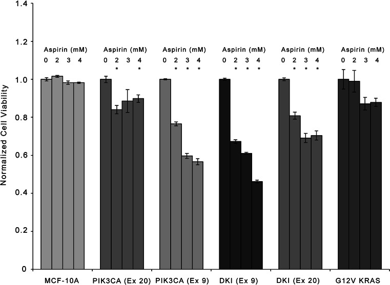
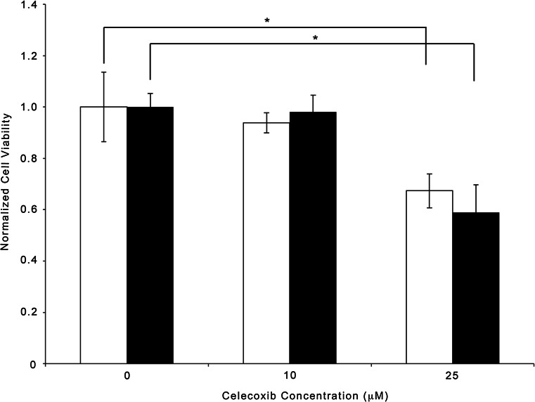
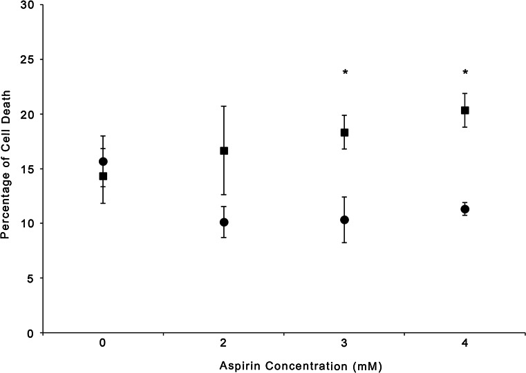
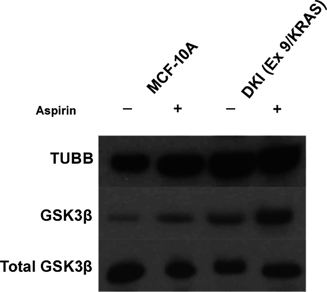
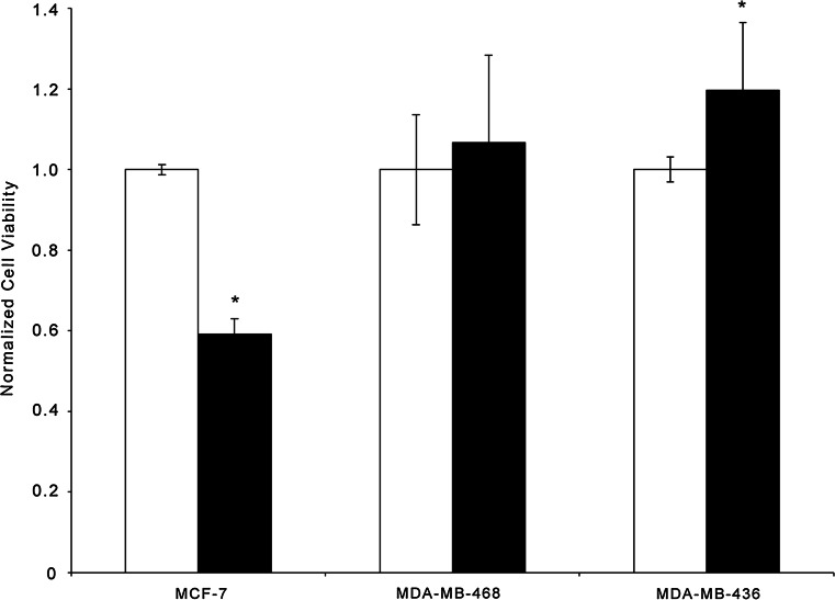
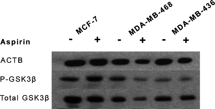
A review of the literature finds that women diagnosed with breast cancer, who were on an aspirin regimen, experienced a decreased risk of distant metastases and death. Several recent studies have reported an improvement in overall survival in colorectal cancer patients who harbored mutations in the oncogene PIK3CA and received a daily aspirin regimen. Breast cancer patients on a daily aspirin regimen experienced decreased risk of distant metastases and death. PIK3CA is the most frequently mutated oncogene in breast cancer, occurring in up to 45 % of all breast cancers. In order to determine if mutations in PIK3CA sensitized breast cancers to aspirin treatment, we employed the use of isogenic cellular clones of the non-tumorigenic, breast epithelial cell line MCF-10A that harbored mutations in either PIK3CA or KRAS or both. We report that mutations in both PIK3CA and KRAS are required for the greatest aspirin sensitivity in breast cancer, and that the GSK3β protein was hyperphosphorylated in aspirin-treated double knockin cells, but not in other clones/treatments. A more modest effect was observed with single mutant PIK3CA, but not KRAS alone. These observations were further confirmed in a panel of breast cancer cell lines. Our findings provide the first evidence that mutations in PIK3CA sensitize breast cancer cells to aspirin.

Keywords: Aspirin; Breast cancer; Drug resistance; Genetics; KRAS; PIK3CA.


Figures

Fig. 1
Fig. 1
Effect of aspirin on parental (MCF-10A), individual knockin mutations of either PIK3CA H1047R, PIK3CA E545K, KRAS G12V, and double mutant knock-ins of KRAS G12Vwith either PIK3CA H1047R or PIK3CA E545K clones of MCF-10A were treated with varying doses of aspirin for 72 h. Parental and clones of MCF-10A were seeded on day 0 in EGF-free media. Following 24 h, the media were replaced with assay media containing 0.2 ng/mL EGF and supplemented with 0–4 mM aspirin. Cells were counted after 72 h. Values for cells treated with 2, 3, and 4 mM aspirin were normalized to the respective 0 mM aspirin value for that particular cell line. Values represent mean ± SEM (*P < 0.05, n = 4)
Fig. 2
Fig. 2
Effect of celecoxib on parental (MCF-10A; white) and double mutant knock-ins of KRAS G12V with PIK3CA E545K (black) clones of MCF-10A were treated with various concentrations of celecoxib for 72 h. Parental MCF-10A and clone of MCF-10A were seeded on day 0 in EGF-free media. Following 24 h, the media were replaced with assay media containing 0.2 ng/mL EGF and supplemented with 0, 10 or 25 µM celecoxib. Cells were counted after 72 h. Cell values for 10 or 25 µM celecoxib were normalized to the respective 0 mM celecoxib value for that particular cell line. Values represent mean ± SD (*P < 0.05, n = 3)
Fig. 3
Fig. 3
Cytotoxic effect of aspirin on parental MCF-10A and MCF-10A DKI (Exon 9/KRAS) cells. Parental (circle) and DKI (square) were seeded on day 0 in EGF-free media for 24 h. The media were then replaced with assay media containing 0.2 ng/mL EGF and supplemented with 0–4 mM aspirin. Cytotoxicity was evaluated after 72 h by measuring uptake of propidium iodide by FACS analysis. Values represent mean ± SD (*P < 0.05, n = 3)
Fig. 4
Fig. 4
Aspirin treatment results in increased phosphorylation of GSK3β in a mutant PIK3CA-dependent manner. Western blot analysis was performed on parental MCF-10A and DKI (Ex 9_KRAS) clones following aspirin treatment against phosphorylated GSK3β (Ser 9) and total GSK3β. TUBB was used as a loading control. Western blot analysis revealed a statistically significant increase (P < 0.05) in the levels of phosphorylated GSK3β following 4 mM aspirin treatment in DKI cells in comparison to non-treated controls and parental MCF-10A cells, regardless of treatment. Blot is representative of three independent experiments
Fig. 5
Fig. 5
Aspirin effect on growth of MCF-7, MDA-MB-468, and MDA-MB-436 cancer cell lines. Cell lines were seeded on day 0. Following 24 h, aspirin (4 mM; black) or no aspirin (white) was added to the assay media. Following 72 h, cells were counted. Cell values for 4 mM aspirin, for each cell line, were normalized to the 0 mM aspirin value for that particular cell line. Cell death due to addition of aspirin was observed in the MCF-7 cell line. MDA-MB-468 and MDA-MB-436 cells did not respond to aspirin treatment. Values represent mean ± SD (*P < 0.05, n = 6)
Fig. 6
Fig. 6
Breast cancer cells expression of GSK3β via Western blot. MCF-7, MDA-MB-468, and MDA-MB-436 cells were seeded on day 0. Next day, the media were replaced, and cells were treated with 4 mM aspirin or no aspirin. After 48 h, media and aspirin were replenished. The next day, cells were harvested in Laemmli buffer, and Western blot was performed to probe for phosphorylated GSK3β (Ser 9) and total GSK3β. ACTB was used as a loading control. Western blot analysis demonstrated that levels of phosphorylated GSK3β following aspirin (4 mM) treatment in MCF-7 cells were slightly increased in comparison to non-treated samples. Figure is representative of three independent experiments

References

    1. What are the key statistics about breast cancer? (2012) http://www.cancer.org/Cancer/BreastCancer/DetailedGuide/breast-cancer-ke...
    1. Fraser DM, Sullivan FM, Thompson AM, McCowan C. Aspirin use and survival after the diagnosis of breast cancer: a population-based cohort study. Br J Cancer. 2014;111(3):623–627. doi: 10.1038/bjc.2014.264. - DOI - PMC - PubMed
    1. Agrawal A, Fentiman IS. NSAIDs and breast cancer: a possible prevention and treatment strategy. Int J Clin Pract. 2008;62(3):444–449. doi: 10.1111/j.1742-1241.2007.01668.x. - DOI - PubMed
    1. Din FV, Valanciute A, Houde VP, Zibrova D, Green KA, Sakamoto K, Alessi DR, Dunlop MG. Aspirin inhibits mTOR signaling, activates AMP-activated protein kinase, and induces autophagy in colorectal cancer cells. Gastroenterology. 2012;142(7):1504–1515. doi: 10.1053/j.gastro.2012.02.050. - DOI - PMC - PubMed
    1. Pasche B, Wang M, Pennison M, Jimenez H. Prevention and treatment of cancer with aspirin: where do we stand? Semin Oncol. 2014;41(3):397–401. doi: 10.1053/j.seminoncol.2014.04.012. - DOI - PMC - PubMed

Publication types