. 2016 Apr 1;594(7):1931-52.
doi: 10.1113/JP271361. Epub 2016 Feb 27.

Long-term exercise-specific neuroprotection in spinal muscular atrophy-like mice

Affiliations

Long-term exercise-specific neuroprotection in spinal muscular atrophy-like mice

Farah Chali et al. J Physiol. .

Abstract

Key points: The real impact of physical exercise parameters, i.e. intensity, type of contraction and solicited energetic metabolism, on neuroprotection in the specific context of neurodegeneration remains poorly explored. In this study behavioural, biochemical and cellular analyses were conducted to compare the effects of two different long-term exercise protocols, high intensity swimming and low intensity running, on motor units of a type 3 spinal muscular atrophy (SMA)-like mouse model. Our data revealed a preferential SMA-induced death of intermediate and fast motor neurons which was limited by the swimming protocol only, suggesting a close relationship between neuron-specific protection and their activation levels by specific exercise. The exercise-induced neuroprotection was independent of SMN protein expression and associated with specific metabolic and behavioural adaptations with notably a swimming-induced reduction of muscle fatigability. Our results provide new insight into the motor units' adaptations to different physical exercise parameters and will contribute to the design of new active physiotherapy protocols for patient care.

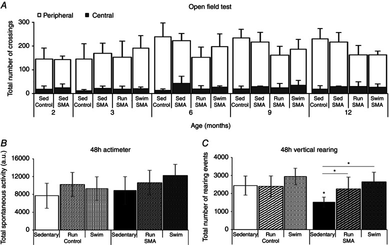
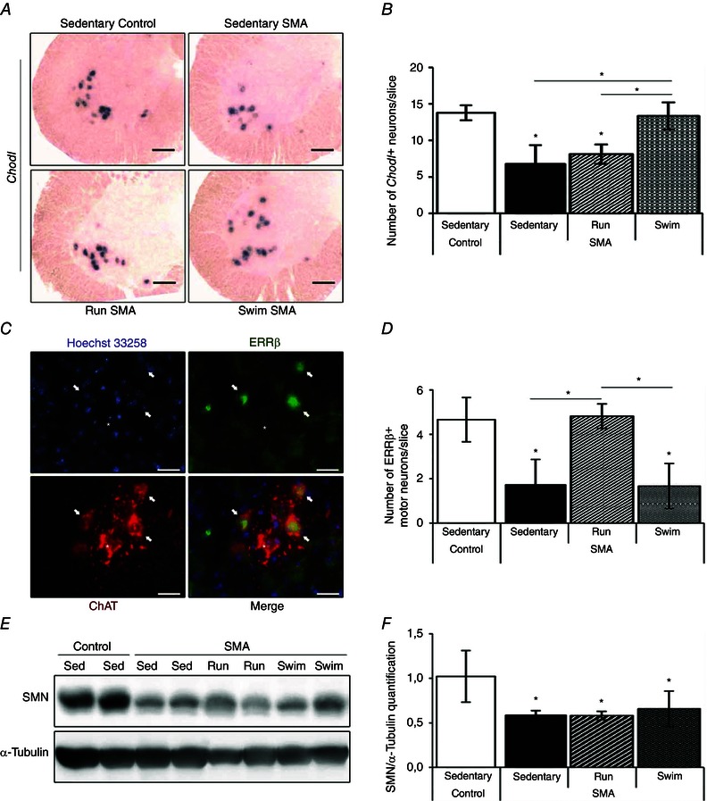
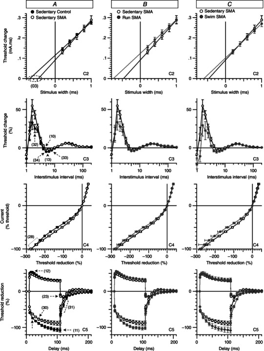
Abstract: Spinal muscular atrophy (SMA) is a group of autosomal recessive neurodegenerative diseases differing in their clinical outcome, characterized by the specific loss of spinal motor neurons, caused by insufficient level of expression of the protein survival of motor neuron (SMN). No cure is at present available for SMA. While physical exercise might represent a promising approach for alleviating SMA symptoms, the lack of data dealing with the effects of different exercise types on diseased motor units still precludes the use of active physiotherapy in SMA patients. In the present study, we have evaluated the efficiency of two long-term physical exercise paradigms, based on either high intensity swimming or low intensity running, in alleviating SMA symptoms in a mild type 3 SMA-like mouse model. We found that 10 months' physical training induced significant benefits in terms of resistance to muscle damage, energetic metabolism, muscle fatigue and motor behaviour. Both exercise types significantly enhanced motor neuron survival, independently of SMN expression, leading to the maintenance of neuromuscular junctions and skeletal muscle phenotypes, particularly in the soleus, plantaris and tibialis of trained mice. Most importantly, both exercises significantly improved neuromuscular excitability properties. Further, all these training-induced benefits were quantitatively and qualitatively related to the specific characteristics of each exercise, suggesting that the related neuroprotection is strongly dependent on the specific activation of some motor neuron subpopulations. Taken together, the present data show significant long-term exercise benefits in type 3 SMA-like mice providing important clues for designing rehabilitation programmes in patients.


Figures

Figure 1
Figure 1. Running‐ and swimming‐based 10‐month training programmes improve muscle resistance to exercise and aerobic capacity in type 3 SMA‐like mice
AD, quantification of creatine kinase activity (A and C) and lactate level (B and D) in the serum of sedentary control and type 3 SMA‐like mice (Control; SMA) at 2 months of age (A and B) after one bout of either running (One Run) or swimming (One Swim) exercise and in the serum of sedentary and 10‐month‐trained control and type 3 SMA‐like mice to running (Trained Run) or swimming (Trained Swim) at 12 months of age (C and D; n = 12 in each group). Data are represented as means ± SD (*P < 0.05).
Figure 2
Figure 2. Running‐ and swimming‐based 10‐month training programmes differentially improve muscle strength and limit muscle fatigability in type 3 SMA‐like mice
A and B, longitudinal measurements of the fore‐ and hindlimb muscle strength (Grip strength test; A) and the forelimb muscle fatigability to low intensity efforts (Grip test; B) of sedentary control mice (Sed Control) compared to sedentary (Sed SMA), running‐trained (Run SMA) and swimming‐trained (Swim SMA) type 3 SMA‐like mice from 2 to 12 months of age. C, longitudinal measurements of the muscle resistance to maximal intensity efforts in swimming pool of sedentary control (Sed Control) and type 3 SMA‐like (Sed SMA) mice compared to swimming‐based trained control (Swim Control) and type 3 SMA‐like (Swim SMA) mice or to running‐based trained type 3 SMA‐like (Run SMA) mice from 2 to 12 months of age (Swimming fatigability test). The minimal sustained water flow was 3 l min−1 and is indicated in the panel by ‘m’. D, measurement of the muscle resistance to maximal intensity efforts in treadmill of sedentary control (Sed Control) and type 3 SMA‐like (Sed SMA) mice compared to swimming‐based trained control (Swim Control) and type 3 SMA‐like (Swim SMA) mice or to running‐based trained type 3 SMA‐like (Run SMA) mice (Running fatigability test). The minimal sustained speed was fixed at 16 m min−1 and is indicated in the panel by ‘m’ (n = 12 in each group). Data are represented as means ± SD (*P < 0.05).
Figure 3
Figure 3. Running‐ and swimming‐based 10‐month training programmes improve motor capacity despite specific adaptations in type 3 SMA‐like mice
A, longitudinal measurements of the exploratory behaviour (Open field test) of sedentary control mice (Sed Control) compared to sedentary (Sed SMA), running‐trained (Run SMA) and swimming‐trained (Swim SMA) type 3 SMA‐like mice from 2 to 12 months of age. B and C, endpoint quantification of the spontaneous activity (B) and vertical rearing events (C) during 48 h (48 h actimeter and 48 h vertical rearing) in sedentary and running‐trained (Run) or swimming‐trained (swim) control and type 3 SMA‐like mice at 12 months of age (n = 12 in each group). Data are represented as means ± SD (*P < 0.05).
Figure 4
Figure 4. Running‐ and swimming‐based 10‐month training programmes induced neuroprotection of motor neuron subpopulations in type 3 SMA‐like mice
A, immunodetection of ChAT‐positive motor neurons in the lumbar spinal cord (L1–L5) of sedentary control mice (left) compared to sedentary (middle left), running‐trained (middle right) and swimming‐trained (right) type 3 SMA‐like mice at 12 months of age (scale bar: 50 μm). BD, quantitative analysis of the number of total, medial and lateral motor neurons per ventral horn (B) and of the absolute number (C) or proportion (D) of motor neurons in small (> 300 μm²), intermediate (300–600 μm²) and large (< 600 μm²) range of cell body area in the spinal cord of sedentary control mice compared to sedentary, running‐trained (Run) and swimming‐trained (Swim) type 3 SMA‐like mice at 12 months of age (n = 8 for each group). Data are represented as means ± SD (*P < 0.05).
Figure 5
Figure 5. Exercise‐induced specific neuroprotection of fast vs. slow motor neuron subpopulations in type 3 SMA‐like mice is SMN independent
A, in situ hybridization on Chondrolectin mRNA (Chodl) in lumbar spinal cords of sedentary control (top left) and type 3 SMA‐like mice (top right) compared to running‐trained (bottom left) and swimming‐trained (bottom right) type 3 SMA‐like mice at 12 months of age (scale bar: 100 μm). B, quantitative analysis of the number of Chodl‐positive neurons in the ventral horn of lumbar spinal cords of sedentary control compared to sedentary, running‐trained (Run) and swimming‐trained (Swim) type 3 SMA‐like mice at 12 months of age (n = 4 in each group). C, immunodetection of the slow‐type motor neuron marker oestrogen‐related receptor β (ERRβ, green) in the nucleus (Hoechst 33258, blue) of ChAT‐positive motor neurons (ChAT, red) in the lumbar spinal cords of sedentary type 3 SMA‐like mice at 12 months of age (scale bar: 25 μm). D, quantitative analysis of the number of ERRβ‐positive motor neurons in the ventral horn of lumbar spinal cords of sedentary control mice compared to sedentary, running‐trained (Run) or swimming‐trained (Swim) type 3 SMA‐like mice at 12 months of age (n = 4 in each group). E and F, Western blot analysis (E) and quantification (F) of SMN protein expression in the ventral lumbar spinal cord of sedentary control and type 3 SMA‐like mice (Sed) compared to running‐trained (Run) or swimming‐trained (Swim) type 3 SMA‐like mice at 12 months of age (n = 4 in each group). Data are represented as means ± SD (*P < 0.05).
Figure 6
Figure 6. Running‐ and swimming‐based 10‐month training programmes reduce efficiently the neuromuscular junction defects in type 3 SMA‐like mice
A–E, motor end‐plate labelling with α‐bungarotoxin (green) and anti‐neurofilament plus anti‐synaptophysin antibodies (NF+Synaptophysin, red), representing a typical pretzel‐shaped (A, left) and a fragmented NMJ (A, right), in the soleus of sedentary control mice (B), sedentary type 3 SMA‐like mice (C) and running‐trained (D) or swimming‐trained type 3 SMA‐like mice (E) at 12 months of age (scale bars: 5 μm for A and 10 μm for BE). F and G, quantification of the neuromuscular junction fragmentation (F) and area (G) in the soleus, plantaris and tibialis muscles from sedentary control mice compared to sedentary, running‐trained (Run) or swimming‐trained (Swim) type 3 SMA‐like mice at 12 months of age (n = 4 in each group). Data are represented as means ± SD (*P < 0.05).
Figure 7
Figure 7. Exercise‐specific adaptations of skeletal muscles in type 3 SMA‐like mice
A, haematoxylin–eosin staining of plantaris of sedentary control mice (top left) compared to sedentary (top right), running‐trained (bottom left) and swimming‐trained (bottom right) type 3 SMA‐like mice at 12 months of age (scale bar: 25 μm). BD, quantification of the cross‐sectional area of myofibres from soleus (B), plantaris (C) and tibialis (D) muscles of sedentary control mice compared to sedentary, running‐trained (Run) and swimming‐trained (Swim) type 3 SMA‐like mice at 12 months of age (n = 4 in each group). E, immunodetection of type IIa myosin heavy chain (MyHC) in the plantaris of sedentary control mice (top left) compared to sedentary (top right), running‐trained (bottom left) and swimming‐trained (bottom right) type 3 SMA‐like mice at 12 months of age (scale bar: 50 μm). F, analysis of adult (i.e. I, II, IIa and IIx/IIb) MyHC isoforms typology of the soleus, plantaris and tibialis of sedentary control mice compared to sedentary, running‐trained (Run) and swimming‐trained (Swim) type 3 SMA‐like mice at 12 months of age (n = 4 in each group). Data are represented as means ± SD (*P < 0.05).
Figure 8
Figure 8. Running‐ and swimming‐based 10‐month training programmes counteract the neuromuscular excitability defect in a type 3 SMA‐like mice
AC, superimposed excitability waveforms obtained in vivo by stimulating the tibial branch of the sciatic nerve and recording the CMAP from plantar muscle in sedentary control mice compared to sedentary type 3 SMA‐like mice (A) and in sedentary type 3 SMA‐like mice compared to either running‐trained (B) or swimming‐trained (C) type 3 SMA‐like mice. The significant differences in excitability parameters between sedentary control and SMA mice are indicated by numbers and dashed lines or arrows. C2, strength–duration relationship. C3, recovery cycle. C4, current–threshold relationship. C5, threshold electrotonus (subthreshold conditioning depolarizing and hyperpolarizing current set to 40%). Data are represented as means ± SD of 4–8 animals per group (*P < 0.05).

References

    1. Bacskai T, Rusznak Z, Paxinos G & Watson C (2014). Musculotopic organization of the motor neurons supplying the mouse hindlimb muscles: a quantitative study using Fluoro‐Gold retrograde tracing. Brain Struct Funct 219, 303–321. - PubMed
    1. Biondi O, Branchu J, Sanchez G, Lancelin C, Deforges S, Lopes P, Pariset C, Lecolle S, Cote J, Chanoine C & Charbonnier F (2010). In vivo NMDA receptor activation accelerates motor unit maturation, protects spinal motor neurons, and enhances SMN2 gene expression in severe spinal muscular atrophy mice. J Neurosci 30, 11288–11299. - PMC - PubMed
    1. Biondi O, Grondard C, Lecolle S, Deforges S, Pariset C, Lopes P, Cifuentes‐Diaz C, Li H, della Gaspera B, Chanoine C & Charbonnier F (2008). Exercise‐induced activation of NMDA receptor promotes motor unit development and survival in a type 2 spinal muscular atrophy model mouse. J Neurosci 28, 953–962. - PMC - PubMed
    1. Bladen CL, Thompson R, Jackson JM, Garland C, Wegel C, Ambrosini A, Pisano P, Walter MC, Schreiber O, Lusakowska A, Jedrzejowska M, Kostera‐Pruszczyk A, van der Pol L, Wadman RI, Gredal O, Karaduman A, Topaloglu H, Yilmaz O, Matyushenko V, Rasic VM, Kosac A, Karcagi V, Garami M, Herczegfalvi A, Monges S, Moresco A, Chertkoff L, Chamova T, Guergueltcheva V, Butoianu N, Craiu D, Korngut L, Campbell C, Haberlova J, Strenkova J, Alejandro M, Jimenez A, Ortiz GG, Enriquez GV, Rodrigues M, Roxburgh R, Dawkins H, Youngs L, Lahdetie J, Angelkova N, Saugier‐Veber P, Cuisset JM, Bloetzer C, Jeannet PY, Klein A, Nascimento A, Tizzano E, Salgado D, Mercuri E, Sejersen T, Kirschner J, Rafferty K, Straub V, Bushby K, Verschuuren J, Beroud C & Lochmuller H (2014). Mapping the differences in care for 5,000 spinal muscular atrophy patients, a survey of 24 national registries in North America, Australasia and Europe. J Neurol 261, 152–163. - PubMed
    1. Boerio D, Greensmith L & Bostock H (2009). Excitability properties of motor axons in the maturing mouse. J Peripher Nerv Syst 14, 45–53. - PubMed

Publication types

MeSH terms

Substances