Review
. 2016 Jul 15;594(14):3993-4008.
doi: 10.1113/JP271827. Epub 2016 May 5.

Cyclic nucleotide regulation of cardiac sympatho-vagal responsiveness

Affiliations
Review

Cyclic nucleotide regulation of cardiac sympatho-vagal responsiveness

Dan Li et al. J Physiol. .

Abstract

Cyclic adenosine monophosphate (cAMP) and cyclic guanosine monophosphate (cGMP) are now recognized as important intracellular signalling molecules that modulate cardiac sympatho-vagal balance in the progression of heart disease. Recent studies have identified that a significant component of autonomic dysfunction associated with several cardiovascular pathologies resides at the end organ, and is coupled to impairment of cyclic nucleotide targeted pathways linked to abnormal intracellular calcium handling and cardiac neurotransmission. Emerging evidence also suggests that cyclic nucleotide coupled phosphodiesterases (PDEs) play a key role limiting the hydrolysis of cAMP and cGMP in disease, and as a consequence this influences the action of the nucleotide on its downstream biological target. In this review, we illustrate the action of nitric oxide-CAPON signalling and brain natriuretic peptide on cGMP and cAMP regulation of cardiac sympatho-vagal transmission in hypertension and ischaemic heart disease. Moreover, we address how PDE2A is now emerging as a major target that affects the efficacy of soluble/particulate guanylate cyclase coupling to cGMP in cardiac dysautonomia.


Figures

Figure 1
Figure 1. Distribution of neuronal nitric oxide synthase (nNOS) protein in the heart and the disease progression from healthy state to heart failure where changes in nNOS may contribute to the aetiology of disease
Modified from Zhang et al. (2014) with permission.
Figure 2
Figure 2. Calcium signalling was significantly enhanced in sympathetic neurons from spontaneously hypertensive rats
A, bright field and immunofluorescence staining image of a cultured cardiac sympathetic neuron derived from a stellate ganglion that was stained with the catecholamine neuronal marker tyrosine hydroxylase (TH, red) and co‐stained with the nuclear marker 4′,6‐diamidino‐ 2‐phenylindole (DAPI, blue). Scale bar represents 20 μm. B, current density–voltage relationship curve of the neuronal calcium current (I Ca) demonstrating significantly larger I Ca at multiple voltages in stellate neurons from spontaneously hypertensive rats (SHRs, n = 8) when compared with Wistar‐Kyoto (WKY) rat controls (n = 8). Ca, typical fluorescence ratio profile of single sympathetic neurons after exposure to high KCl. b, pseudocolour‐coded ratio images of Fura‐2‐loaded neuron were obtained by conventional fluorescence microscopy. Ca2+ concentrations were colour‐coded with a basal Ca2+ concentration in blue and a high Ca2+ concentration in red. c, statistical data showed peak increase in intracellular free calcium concentration ([Ca2+]i) on high K+ exposure from SHRs compared with WKY rats of the same age. *P < 0.05, **P < 0.01, t test. P < 0.05, †† P < 0.01, one‐way ANOVA. Modified from Li et al. (2012, 2015) and Lu et al. (2015) with permission.
Figure 3
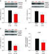
Figure 3. Protein expression of CAPON, nNOS, β1‐sGC and cGMP concentration was reduced in sympathetic ganglia from spontaneously hypertensive rats
AD, representative Western blot and group mean data showing a significant reduction in CAPON (A), nNOS (B) and β1‐sGC (C) protein expression relative to β‐actin in stellate ganglia from 4‐week‐old spontaneously hypertensive rats (SHR) compared with age matched Wistar–Kyoto (WKY) controls. D, tissue levels of cGMP in the stellate ganglia from the young SHRs were significantly lower than that measured in the WKY rats. (*P < 0.05, **P < 0.01, unpaired t test.) Modified from Li et al. (2013) and Lu et al. (2015) with permission.
Figure 4
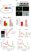
Figure 4. Overexpress of CAPON in sympathetic neuron rescues impaired NO‐cGMP pathway in spontaneously hypertensive rats
A, map of adenoviral vector construct containing a noradrenergic neuron‐specific promoter (PRSx8), NOS1‐AP (neuronal nitric oxide synthase adaptor protein [CAPON]) gene, and red mCherry fluorescent protein (Ad.PRSx8‐mCherry/CAPON). As a control, the same construct without CAPON gene insert was used (Ad.PRSx8‐mCherry). Ba, representative Western blot showing CAPON expression (74 kDa) in stellate ganglia from 4‐week‐old SHR 3 days after transduction with Ad.PRSx8‐mCherry/CAPON (Ad.CAPON) and Ad.PRSx8‐mCherry (empty). b, co‐immunostaining of stellate neurons with anti‐tyrosine hydroxylase (TH, green) and mCherry (red) tagged viral construct showed viral transduction in sympathetic neurons. Nuclear staining with DAPI is in blue. Scale bar, 25 μm. C, gene transfer of CAPON (Ad.CAPON) significantly increased NOS activity in stellate ganglia from SHR when compared with the empty vector control. The specific nNOS inhibitor, S2, N‐[(4S)‐4‐amino‐5‐[(2‐aminoetyl) amino] pentyl]‐N′‐nitroguanidine (AAAN, 10 μmol L−1) normalized the difference in NOS activity after transduction with CAPON or the empty vector control (*P < 0.05, ANOVA). D, cGMP concentration in SHR stellate ganglia tissue was significantly enhanced by Ad.CAPON transduction when compared with empty controls (*P < 0.05, unpaired t test). E, current density–voltage relationship curve of the neuronal calcium current (I Ca) demonstrating attenuation of I Ca at multiple voltages in CAPON‐overexpressing stellate neurons from 4‐week‐old SHR when compared with cells transduced with empty virus. Inset images: representative fluorescence (mCherry) and bright field images of single stellate neuron transduced with Ad.CAPON with a patch pipette (indicated by a white arrow). F and G, gene transfer with CAPON in SHRs significantly reduced the calcium transient in sympathetic neurons (F) and [3H]noradreanaline (NA) release (G) from isolated double atrial preparations when compared with the empty vector. The atria were stimulated at 5 Hz for 1 min at the 16th (S1) and 40th (S2) minutes. Bath solution was collected by replacing 3 ml Tyrode's solution every 3 min and measured using a liquid scintillation counter. The effect of CAPON gene transfer can be reversed with a specific nNOS inhibitor (AAAN, 10 μmol L−1). Modified from Lu et al. (2015) with permission.
Figure 5
Figure 5
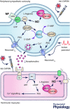
Summary diagram illustrating the potential signal transduction pathways mediated by natriuretic peptides (NP) and nitric oxide (NO)–CAPON in depolarized cardiac stellate neurons
Figure 6
Figure 6. Effect of BNP on the intracellular calcium current and calcium transient in isolated sympathetic neurons from the normal rats
Aa, representative whole cell calcium current density traces obtained with or without 100 nmol L−1 B‐type natriuretic peptide (BNP, 10 min) and after wash out. Currents were evoked by test pulses to −10 mV from a holding potential of −90 mV. b, mean current density–voltage relations in the presence and absence of 100 nmol L−1 BNP. Wash‐off data were only recorded at −10 mV as the quality of the recordings deteriorates over time. *P < 0.05, paired t test, n = 6 neurons. Ba, an example recording of a calcium transient in a single cardiac sympathetic neuron. The neuron was exposed to KCl for 30 s to depolarize it with (S2) or without (S1) BNP. b, statistical data showing that the concentration–effect relationship of BNP (1–250 nmol L−1) changed KCl‐evoked increase in [Ca2+]i expressed as a ratio (%) of S2 compared with S1. **P < 0.01, compared with control, unpaired t test (n = 8–16). C, grouped data showing the time control (a) and with addition of 250 nmol L−1 BNP (b) on [3H]noradrenaline (NA) release from isolated atria. The atria were stimulated at 5 Hz for 1 min at the 16th (S1) and 40th (S2) minute (n = 6 *P < 0.05). Bath solution was collected by replacing 3 ml Tyrode's solution every 3 min and measured in a β‐radiation scintillation counter. D, representative raw data traces (a) and grouped data (b) showing the heart rate responses to sympathetic nerve stimulation (SNS) at 1, 3, 5 and 7 Hz for 30 s with 250 nmol L−1 BNP compared with control. n = 10, *P < 0.05, **P < 0.01, paired t test. Modified from Li et al. (2015) with permission.
Figure 7
Figure 7. Overexpress of PDE2A in sympathetic neurons markedly decreases BNP stimulation of cGMP and enhances the intracellular calcium transient in normal rats
Aa and b, Western blot showing PDE2A.mCherry expression (127 kDa) in transduced empty and Ad.PDE2A stellate ganglia tissue (with anti‐PDE2A antibody). Band optical density was normalized to that of β‐actin (42 kDa). c, effect of 250 nmol L−1 BNP on cGMP concentration in transduced empty and PDE2A stellate ganglia tissue. Ba and b, ratio data traces (a) and statistical data (b) showing 50 mmol L−1 KCl‐evoked intracellular calcium transient ([Ca2+]i) in the transduced empty and Ad.PDE2A cardiac sympathetic neurons. c, group data showing noradrenaline (NA) release during 5 Hz field stimulation was significantly enhanced in the percutaneous gene transfer to right atria with Ad.PDE2A when compared with the empty vector. *P < 0.05, **P < 0.01, unpaired t test. C, diagram illustrating the potential signal transduction pathways mediated by B‐type natriuretic peptide (BNP) in depolarized stellate neurons. BNP binds to the natriuretic peptide receptor A (NPR‐A) and stimulates particulate guanylyl cyclase (pGC). Newly synthesized cGMP activates phosphodiesterase 2 (PDE2) to degrade cGMP, but not cAMP, and thus limits the increase in protein kinase G (PKG) activity. Moreover, PKG inhibits calcium current by phosphorylating voltage‐gated calcium channels (VGCCs) and reduces the intracellular calcium transient. This in turn decreases NA release from synaptic vesicles and reduces the heart rate response to sympathetic stimulation. In addition, elevated intracellular calcium triggers the activation of adenylate cyclase (AC) via calmodulin (CaM) and produces cAMP. cAMP activates cAMP‐dependent protein kinase (PKA), and thus phosphorylation of calcium channel. Modified from Li et al. (2015) with permission.

References

    1. Abramson BL, Ando S, Notarius CF, Rongen GA & Floras JS (1999). Effect of atrial natriuretic peptide on muscle sympathetic activity and its reflex control in human heart failure. Circulation 99, 1810–1815. - PubMed
    1. Ackermann U, Khanna J & Irizawa TG (1988). Atrial natriuretic factor alters autonomic interactions in the control of heart rate in conscious rats. Can J Physiol Pharmacol 66, 930–936. - PubMed
    1. Ashman DF, Lipton R, Melicow MM & Price TD (1963). Isolation of adenosine 3′,5′‐monophosphate and guanosine 3′,5′‐monophosphate from rat urine. Biochem Biophys Res Commun 11, 330–334. - PubMed
    1. Atchison DJ & Ackermann U (1993). The interaction between atrial natriuretic peptide and cardiac parasympathetic function. J Auton Nerv Syst 42, 81–88. - PubMed
    1. Balligand JL, Kelly RA, Marsden PA, Smith TW & Michel T (1993). Control of cardiac muscle cell function by an endogenous nitric oxide signaling system. Proc Natl Acad Sci USA 90, 347–351. - PMC - PubMed

Publication types

LinkOut - more resources