. 2016 May;65(5):1350-61.
doi: 10.2337/db15-1525. Epub 2016 Feb 25.

Bioengineering the Endocrine Pancreas: Intraomental Islet Transplantation Within a Biologic Resorbable Scaffold

Affiliations

Bioengineering the Endocrine Pancreas: Intraomental Islet Transplantation Within a Biologic Resorbable Scaffold

Dora M Berman et al. Diabetes. 2016 May.

Abstract

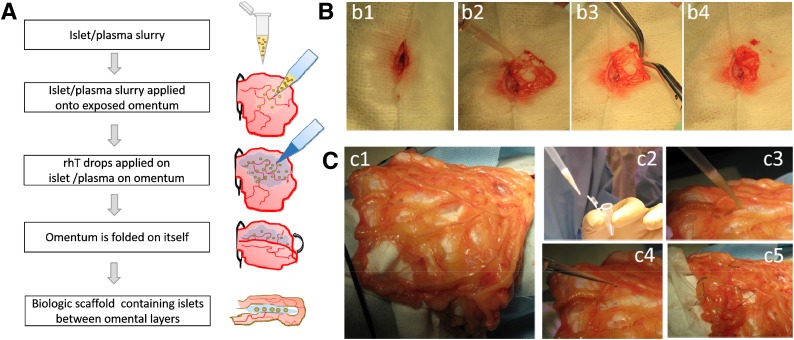
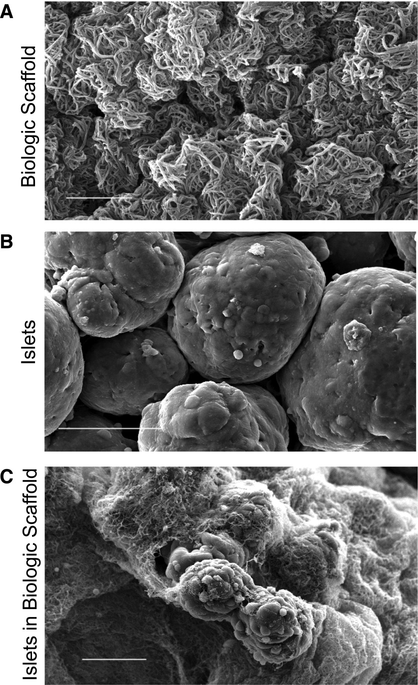
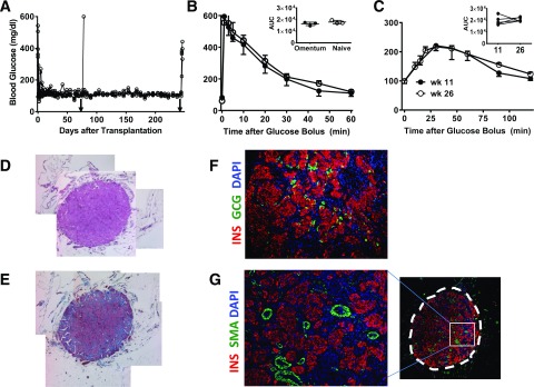
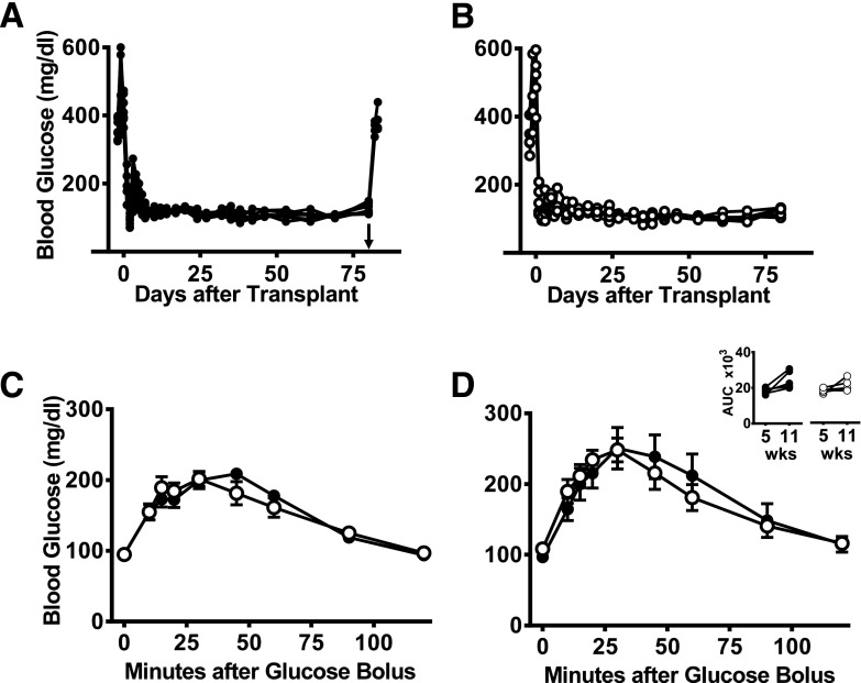
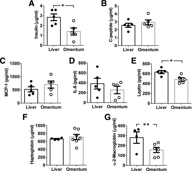
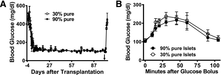
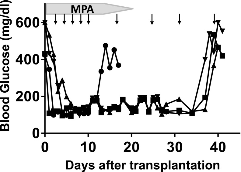
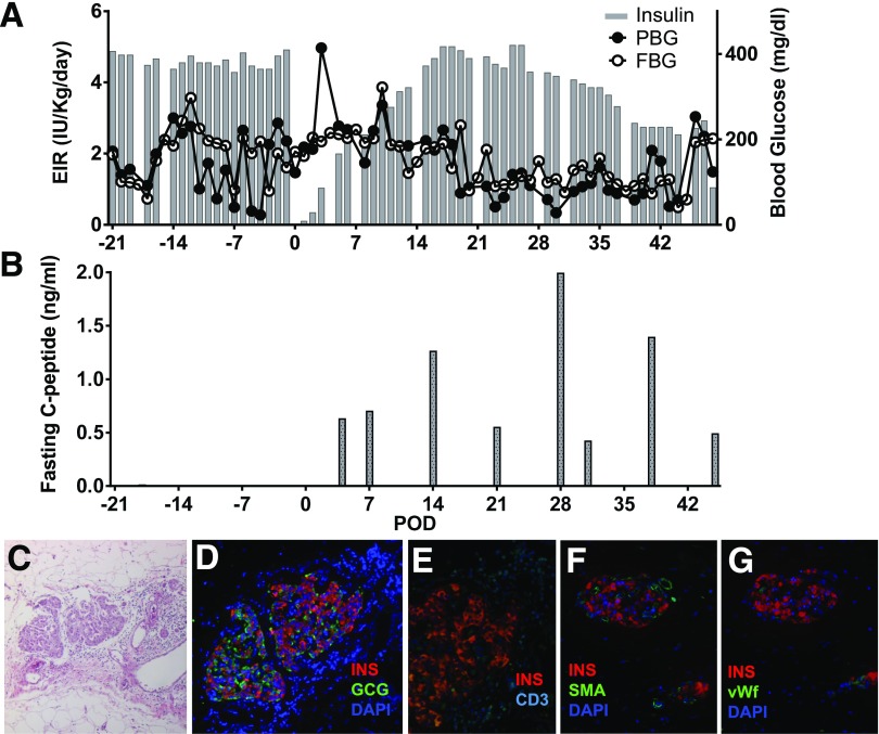
Transplantation of pancreatic islets is a therapeutic option to preserve or restore β-cell function. Our study was aimed at developing a clinically applicable protocol for extrahepatic transplantation of pancreatic islets. The potency of islets implanted onto the omentum, using an in situ-generated adherent, resorbable plasma-thrombin biologic scaffold, was evaluated in diabetic rat and nonhuman primate (NHP) models. Intraomental islet engraftment in the biologic scaffold was confirmed by achievement of improved metabolic function and preservation of islet cytoarchitecture, with reconstitution of rich intrainsular vascular networks in both species. Long-term nonfasting normoglycemia and adequate glucose clearance (tolerance tests) were achieved in both intrahepatic and intraomental sites in rats. Intraomental graft recipients displayed lower levels of serum biomarkers of islet distress (e.g., acute serum insulin) and inflammation (e.g., leptin and α2-macroglobulin). Importantly, low-purity (30:70% endocrine:exocrine) syngeneic rat islet preparations displayed function equivalent to that of pure (>95% endocrine) preparations after intraomental biologic scaffold implantation. Moreover, the biologic scaffold sustained allogeneic islet engraftment in immunosuppressed recipients. Collectively, our feasibility/efficacy data, along with the simplicity of the procedure and the safety of the biologic scaffold components, represented sufficient preclinical testing to proceed to a pilot phase I/II clinical trial.


Figures

Figure 1
Figure 1
Intraomental islet implantation within a biologic scaffold. A: Schematic diagram of the transplant procedure. B: Procedure in rat. C: Procedure in NHP. After midline laparotomy (b1), the omentum is gently exteriorized and opened (b2 and c1). The islet graft, resuspended in autologous plasma (c2), is gently distributed onto the omentum (b3 and c3). Recombinant human thrombin is added onto the islets on the omental surface to induce gel formation (c4), and then the omentum is folded to increase the contact of the graft to the vascularized omentum (b4 and c5). Nonresorbable stitches were placed on the far outer margins of the graft in the NHP (c5) for easier identification of the graft area at the time of graft removal.
Figure 2
Figure 2
Scanning electronic micrograph of the biologic scaffold in vitro. A: Plasma/thrombin mix. Fibrin polymerizes forming an intricate three-dimensional network (bar = 5 µm). B: Untreated human islet cell surface in culture medium (bar = 50 µm). C: Human islets embedded within the biologic scaffold. The polymerized fibrin forms an orthomorphic matrix around the islet surface (bar = 50 µm).
Figure 3
Figure 3
Intraomental islets transplanted into biologic scaffolds restore normoglycemia in diabetic rats. A: Nonfasting blood glucose levels in diabetic rats (n = 7; 173.4 ± 91 g body wt) transplanted with 3,000 IEQ (17,338 ± 881 IEQ/kg) onto the omentum showing prompt reversal of diabetes and hyperglycemia after removal of the omental graft (arrowhead) on POD 74 (n = 1) or 240 (n = 4). B: Glycemic profile during IVGTT performed in selected animals (n = 3) 2 months after transplant compared with that of naïve animals (n = 3). Values shown are mean ± SD. Inset shows area under the curve (AUC) (mg × min × dL−1) for each group. C: Glycemic profile during OGTT performed in transplanted animals (n = 5) at 11 (●) and 26 (○) weeks after transplantation. Inset shows AUC (mg × min × dL−1) during the glucose challenge. DG: Representative histopathologic pattern of intraomental islet grafts. Sections were obtained from an intraomental islet graft explanted on POD 76. D: Hematoxylin-eosin staining. E: Masson trichrome staining. F and G: Immunofluorescence microscopy of a section stained with anti-insulin (INS) (red fluorescence), anti-GCG antibody (green fluorescence) (F), anti-SMA (green fluorescence) (G), and nuclear dye DAPI (blue fluorescence). The box indicates the area of the graft shown at higher magnification on the left panel. wk, week.
Figure 4
Figure 4
Comparable function of intrahepatic and intraomental islets transplanted into biologic scaffolds. Nonfasting blood glucose levels in diabetic rats receiving a clinically relevant syngeneic islet mass of 1,300 IEQ (∼8,200 IEQ/kg body wt) within an intraomental biologic scaffold (A) (●, n = 7) or into the liver (via the portal vein) (B) (○, n = 5) with islets from the same batch isolation. The groups had an identical time course for reversal of diabetes, and removal of the intraomental biologic scaffold on day 80 posttransplant resulted in return to hyperglycemia (arrowhead in A). Glycemic profile during OGTT performed in all transplanted animals 5 (C) or 11 weeks (D) after transplantation. Inset shows AUC (mg × min × dL−1) during the glucose challenge for each group. wks, weeks.
Figure 5
Figure 5
Biomarkers detected in the serum of rat recipients of intraomental biologic scaffold and intrahepatic syngeneic islets. Aliquots of 1,300 IEQ from the same syngeneic donor rat islet batch were transplanted in parallel either within the intraomental biologic scaffold (omentum [○]) or the intrahepatic site (liver [●]). Blood samples were collected from indwelling JVC for detection of biomarker levels in circulation. Data presented are mean ± SEM (n = 4–7 per time point). A and B: Metabolic markers assessed at 1 h posttransplant. A: Insulin in μg/mL (*P = 0.018). B: C-peptide in μg/mL. Inflammation markers assessed 24 h posttransplant: MCP-1/CCL2 in pg/mL (C), IL-6 in pg/mL (D), leptin in pg/mL (*P = 0.013) (E), haptoglobin in μg/mL (F), and α2-macroglobulin in μg/mL (**P < 0.03) (G).
Figure 6
Figure 6
Intraomental transplantation of islets with high and low purity into diabetic rats. A: Nonfasting blood glucose levels in diabetic rats transplanted with clinically relevant mass of 2,000 IEQ syngeneic islets with >95% purity (n = 3) (167.3 ± 1.5 g body wt [11,853 ± 109 IEQ/kg]) or with 30% purity (n = 3) (170.3 ± 10.5 g body wt [11,771 ± 725 IEQ/kg]) onto the omentum. Removal of the omental graft >100 days after transplantation (arrowhead) resulted in return to hyperglycemia. B: Glycemic profile during oral glucose tolerance test performed in animals transplanted with high-purity and low-purity islet preparations 70 days after transplantation.
Figure 7
Figure 7
The intraomental biologic scaffold supports the engraftment of allogeneic islets under systemic immunosuppression in diabetic rats. A fully MHC-mismatched allogeneic rat transplant combination in which diabetic female Lewis rat (RT1l) (n = 4) received 3,000 IEQ WF rat islets (RT1u) in the intraomental biologic scaffold under a protocol of clinically relevant immunosuppressive agents consisting of lymphodepletion induction with anti-lymphocyte serum (0.5 mL i.p. on day −3) and maintenance with mycophenolic acid (MPA) (20 mg/kg/day for days 0–14, then tapered by one-quarter of the dose every 2 days until day 20) and CTLA4Ig (10 mg/kg i.p. on days 0, 2, 4, 6, 8, and 10 and weekly thereafter; abatacept) (arrows). Nonfasting glycemic values for each animal during the follow-up are presented. Graft rejection was defined as return to hyperglycemic state. Each symbol represents an individual animal.
Figure 8
Figure 8
Intraomental allogeneic islet transplantation in a diabetic nonhuman primate. A diabetic cynomolgus monkey received 9,347 IEQ/kg allogeneic islets in the omentum under the cover of clinically relevant immunosuppression therapy. A: Exogenous insulin requirement (EIR) (IU/kg/day), FBG (mg/dL), and PBG. B: Fasting C-peptide (ng/mL) levels measured in the animal over the follow-up period. CG: Histopathologic pattern of intraomental islet graft on day 49 posttransplant. C: Hematoxylin-eosin staining. D: Immunofluorescence microscopy for the evaluation of immunoreactivity for insulin (INS) (red), GCG (green), and nuclear dye (DAPI) (blue). E: Immunofluorescence for insulin (red) and CD3+ T cells (CD3) (cyan). F and G: Intrainsular neovasculogenesis. F: Immunofluorescence for insulin (red), vascular structure (SMA) (green), and DAPI (blue). G: Immunofluorescence microscopy for insulin (red), endothelial cells (vWF) (green), and DAPI (blue).

References

    1. Piemonti L, Pileggi A. 25 years of the Ricordi Automated Method for islet isolation. CellR4 2013;1:8–22 - PMC - PubMed
    1. Pileggi A, Ricordi C, Kenyon NS, et al. . Twenty years of clinical islet transplantation at the Diabetes Research Institute--University of Miami. Clin Transpl 2004;177–204 - PubMed
    1. Alejandro R, Cutfield RG, Shienvold FL, et al. . Natural history of intrahepatic canine islet cell autografts. J Clin Invest 1986;78:1339–1348 - PMC - PubMed
    1. Bennet W, Groth CG, Larsson R, Nilsson B, Korsgren O. Isolated human islets trigger an instant blood mediated inflammatory reaction: implications for intraportal islet transplantation as a treatment for patients with type 1 diabetes. Ups J Med Sci 2000;105:125–133 - PubMed
    1. Moberg L, Johansson H, Lukinius A, et al. . Production of tissue factor by pancreatic islet cells as a trigger of detrimental thrombotic reactions in clinical islet transplantation. Lancet 2002;360:2039–2045 - PubMed

MeSH terms