. 2016 Feb 25:9:9.
doi: 10.1186/s13048-016-0219-x.

Progesterone acts via the progesterone receptor to induce adamts proteases in ovarian cancer cells

Affiliations

Progesterone acts via the progesterone receptor to induce adamts proteases in ovarian cancer cells

Maíra A Lima et al. J Ovarian Res. .

Abstract

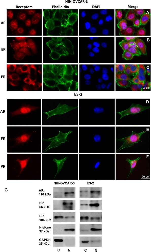
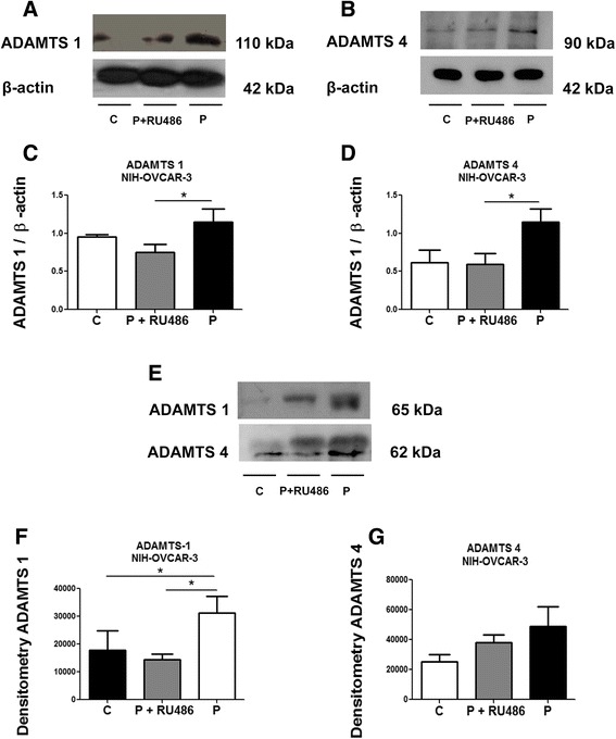
Background: Ovarian carcinomas, usually associated with sex hormones dysregulation, are the leading cause of gynecological neoplastic death. In normal ovaries, hormones play a central role in regulating cell proliferation, differentiation, and apoptosis. On the other hand, hormonal alterations also play a variety of roles in cancer. Stimulation by sex hormones potentially affects gene expression, invasiveness, cell growth and angiogenesis. Proteases of the "a disintegrin and metalloproteinase with thrombospondin motifs" (ADAMTS) family are secreted by different cell types and become involved in collagen processing, cleavage of the proteoglycan matrix, and angiogenesis. We evaluated whether sex hormones affect ADAMTS 1 and 4 expression in ovarian cancer cells.

Methods: We analysed mRNA and protein levels in human ovarian tumor cells with different degrees of malignancy, NIH-OVCAR-3 and ES-2, that were treated or not with estrogen, testosterone and progesterone.

Results: Our results suggest that progesterone increases ADAMTS protein and mRNA levels in the lysates from ES-2 cells, and it increases ADAMTS protein in the lysates and conditioned media from NIH-OVCAR-3. Progesterone effects were reversed by RU486 treatment.

Conclusion: We conclude that progesterone acts via the progesterone receptor to modulate ADAMTS 1 and 4 levels in ovarian cancer cell lines.


Figures

Fig. 1
Fig. 1
Distribution of AR, ER and PR in NIH-OVCAR-3 and ES-2 cells. AR, ER and PR (red), nuclei (blue) and actin (green). Merge dots in magenta indicate the colocalization of the receptors with the nucleus. In NIH-OVCAR-3 cells a Androgen receptor and b Estrogen receptor are mostly in the nucleus, but also appears in cytoplasm. c Progesterone receptor is mostly found in the cell cytoplasm. In ES-2 cells d Androgen receptor and e Estrogen receptor are also mainly in the cell nucleus. f Progesterone receptor is located in cytoplasm and nucleus. g Immunoblot of NIH-OVCAR-3 and ES-2 cell fractions for receptors. In NIH-OVCAR-3 cells ER and AR are present in the nucleus, PR is present in both compartments, but mostly in the cytosolic fraction. ES-2 cells present most of the AR and ER in the nuclear fraction and PR is preferentially in the cytosolic compartment. Scale bar: 20 μm. (C = cytosolic fraction, N = nuclear fraction)
Fig. 2
Fig. 2
Cell viability and ADAMTS mRNA expression in NIH-OVCAR-3 and ES-2 cell lines, after hormone treatment. a and b MTT viability assay of NIH-OVCAR-3 and ES-2 cell lines treated with 30 nM estrogen, 1 μM progesterone or 100 nM testosterone for 24 h compared to control. c and d ADAMTS 1 and 4 mRNA expression assessed by qPCR in the lysates of NIH-OVCAR-3 cells. GAPDH was used as normalizer. e and f ADAMTS 1 and 4 mRNA expression assessed by qPCR in the cell lysates of ES-2 cells. GAPDH was used as normalizer. Differences among groups were considered statistically significant when p ≤ 0.05. Experiments were performed in triplicate
Fig. 3
Fig. 3
ADAMTS 1 and 4 protein expression of NIH-OVCAR-3 cell lysates and conditioned medium, after hormone treatment. a Representative Western blot of ADAMTS 1 and 4 in the lysates of NIH-OVCAR-3 cells treated with 30 nM estrogen (E), 1 μM progesterone (P), or 100 nM testosterone (T), compared to control (C). β-actin was used as loading control. b and c ADAMTS 1 and 4 protein expression in the lysates of NIH-OVCAR-3 cells. d Representative Western blot of ADAMTS 1 and 4 in the conditioned medium from NIH-OVCAR-2 cells. e and f ADAMTS 1 and 4 protein expression in the conditioned medium from NIH-OVCAR-3 cells. Conditioned medium equal sample loading was confirmed by Pounceau S staining (Additional file 1: Fig. S1a). Differences among groups were considered statistically significant when p ≤ 0.05. Experiments were performed in triplicate
Fig. 4
Fig. 4
Progesterone induces increase of ADAMTS 1 and 4 in cell lysates and conditioned medium of ES-2 cell. a and c Representative Western blot of ADAMTS 1 and 4 in the lysates of ES-2 cells treated with 30 nM estrogen (E), 1 μM progesterone (P) or 100 nM testosterone (T), compared to control (C). β-actin was used as loading control. b and d ADAMTS 1 and 4 protein expression in the lysates of ES-2 cells. e Representative Western blot of ADAMTS 1 and 4 in the conditioned medium from ES-2 cells. f and g ADAMTS 1 and 4 protein expression in the conditioned medium from ES-2 cells. Conditioned medium equal sample loading was confirmed by Pounceau S staining (Additional file 1: Fig. S1b). Differences among groups were considered statistically significant when p ≤ 0.05. Experiments were performed in triplicate
Fig. 5
Fig. 5
RU486 can rescue the increase of ADAMTS 1 induced by progesterone in NIH-OVCAR-3 conditioned medium. a and b Representative Western blot of ADAMTS 1 and 4 in the lysates of NIH-OVCAR-3 cells treated with progesterone (P) or progesterone and RU486 (P + RU486) compared to control (C). c and d ADAMTS 1 and 4 protein expression in the lysate of NIH-OVCAR-3 cells. e Representative Western blot of ADAMTS 1 and 4 in the conditioned medium from NIH-OVCAR-3 cells. f and g ADAMTS 1 and 4 protein expression in conditioned medium of NIH-OVCAR-3. Conditioned medium equal sample loading was confirmed by Pounceau S staining (Additional file 1: Fig. S1c). Differences among groups were considered statistically significant when p ≤ 0.05. Experiments were performed in triplicate
Fig. 6
Fig. 6
RU486 can rescue the increase of ADAMTS 1 and 4 induced by progesterone in ES-2 cell lysates but not in the conditioned medium. a and b Representative Western blot of ADAMTS 1 and 4 in the lysates of ES-2 cells treated with progesterone (P) or progesterone and RU486 (P + RU486) compared to control (C). c and d ADAMTS 1 and 4 protein expression in the lysates of ES-2 cells. e Representative Western blot of ADAMTS 1 and 4 in the conditioned medium from ES-2 cells treated as above. f and g ADAMTS 1 and 4 protein expression in the conditioned medium from ES-2 cells. Conditioned medium equal sample loading was confirmed by Pounceau S staining (Additional file 1: Fig. S1d). Differences among groups were considered statistically significant when p ≤ 0.05. Experiments were performed in triplicate

References

    1. Pecorelli S, Favalli G, Zigliani L, Odicino F. Cancer in women. Int J Gynaecol Obstet. 2003;82:369–79. doi: 10.1016/S0020-7292(03)00225-X. - DOI - PubMed
    1. Ween MP, Oehler MK, Ricciardelli C. Role of versican, hyaluronan and CD44 in ovarian cancer metastasis. Int J Mol Sci. 2011;12:1009–29. doi: 10.3390/ijms12021009. - DOI - PMC - PubMed
    1. Lukanova A, Kaaks R. Endogenous hormones and ovarian cancer: epidemiology and current hypotheses. Cancer Epidemiol Biomarkers Prev. 2005;14:98–107. - PubMed
    1. Judd HL, Judd GE, Lucas WE, Yen SS. Endocrine function of the postmenopausal ovary: concentration of androgens and estrogens in ovarian and peripheral vein blood. J Clin Endocrinol Metab. 1974;39:1020–4. doi: 10.1210/jcem-39-6-1020. - DOI - PubMed
    1. Wang PH, Chang C. Androgens and ovarian cancers. Eur J Gynaecol Oncol. 2004;25:157–63. - PubMed

Publication types