. 2016 Mar;139(Pt 3):765-81.
doi: 10.1093/brain/awv393.

EPG5-related Vici syndrome: a paradigm of neurodevelopmental disorders with defective autophagy

Affiliations

EPG5-related Vici syndrome: a paradigm of neurodevelopmental disorders with defective autophagy

Susan Byrne et al. Brain. 2016 Mar.

Abstract

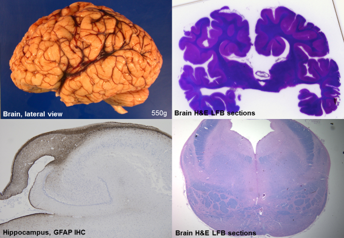
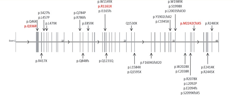
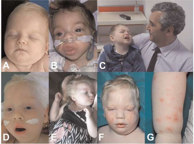
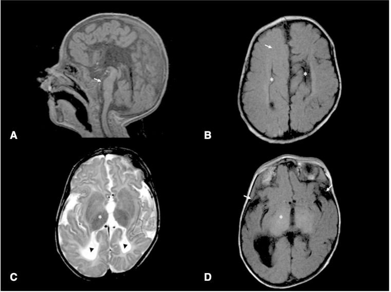
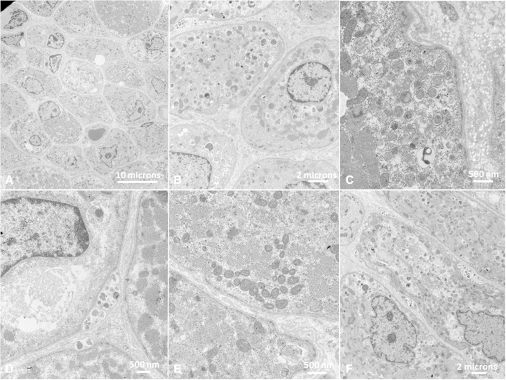
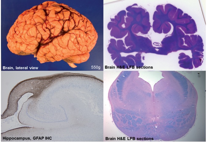
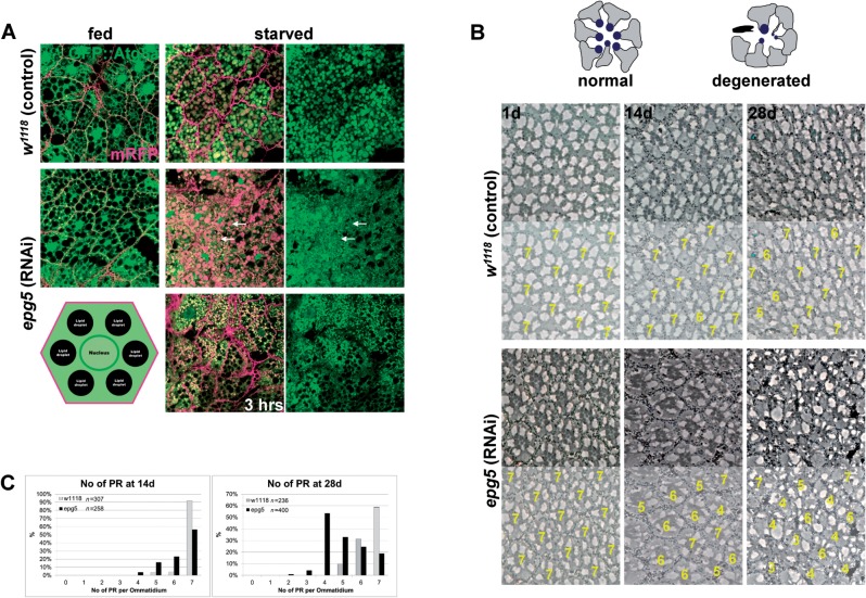
Vici syndrome is a progressive neurodevelopmental multisystem disorder due to recessive mutations in the key autophagy gene EPG5. We report genetic, clinical, neuroradiological, and neuropathological features of 50 children from 30 families, as well as the neuronal phenotype of EPG5 knock-down in Drosophila melanogaster. We identified 39 different EPG5 mutations, most of them truncating and predicted to result in reduced EPG5 protein. Most mutations were private, but three recurrent mutations (p.Met2242Cysfs*5, p.Arg417*, and p.Gln336Arg) indicated possible founder effects. Presentation was mainly neonatal, with marked hypotonia and feeding difficulties. In addition to the five principal features (callosal agenesis, cataracts, hypopigmentation, cardiomyopathy, and immune dysfunction), we identified three equally consistent features (profound developmental delay, progressive microcephaly, and failure to thrive). The manifestation of all eight of these features has a specificity of 97%, and a sensitivity of 89% for the presence of an EPG5 mutation and will allow informed decisions about genetic testing. Clinical progression was relentless and many children died in infancy. Survival analysis demonstrated a median survival time of 24 months (95% confidence interval 0-49 months), with only a 10th of patients surviving to 5 years of age. Survival outcomes were significantly better in patients with compound heterozygous mutations (P = 0.046), as well as in patients with the recurrent p.Gln336Arg mutation. Acquired microcephaly and regression of skills in long-term survivors suggests a neurodegenerative component superimposed on the principal neurodevelopmental defect. Two-thirds of patients had a severe seizure disorder, placing EPG5 within the rapidly expanding group of genes associated with early-onset epileptic encephalopathies. Consistent neuroradiological features comprised structural abnormalities, in particular callosal agenesis and pontine hypoplasia, delayed myelination and, less frequently, thalamic signal intensity changes evolving over time. Typical muscle biopsy features included fibre size variability, central/internal nuclei, abnormal glycogen storage, presence of autophagic vacuoles and secondary mitochondrial abnormalities. Nerve biopsy performed in one case revealed subtotal absence of myelinated axons. Post-mortem examinations in three patients confirmed neurodevelopmental and neurodegenerative features and multisystem involvement. Finally, downregulation of epg5 (CG14299) in Drosophila resulted in autophagic abnormalities and progressive neurodegeneration. We conclude that EPG5-related Vici syndrome defines a novel group of neurodevelopmental disorders that should be considered in patients with suggestive features in whom mitochondrial, glycogen, or lysosomal storage disorders have been excluded. Neurological progression over time indicates an intriguing link between neurodevelopment and neurodegeneration, also supported by neurodegenerative features in epg5-deficient Drosophila, and recent implication of other autophagy regulators in late-onset neurodegenerative disease.

Keywords: EPG5; Vici syndrome; callosal agenesis; ectopic P granules autophagy protein 5; neurodegeneration; neurodevelopment.


Figures

None
Vici syndrome is a progressive neurodevelopmental multisystem disorder caused by mutations in the autophagy gene EPG5. Byrne et al. characterise the phenotype of 50 affected children, revealing callosal agenesis, cataracts, hypopigmentation, cardiomyopathy, immune dysfunction, developmental delay and microcephaly. Downregulation of epg5 in Drosophila results in autophagic abnormalities and progressive neurodegeneration.
Figure 1
Figure 1
Distribution of disease-causing mutations in EPG5. The EPG5 gene is represented, with the 44 exons depicted as grey vertical bars (exons 1, 10, 25, 35 and 44 are numbered for orientation). The exon position of Vici-causing mutations are indicated by the arrows, with the mutation details listed at the arrow tail. Mutations found in two or more unrelated patients due to possible founder effects are coloured red. EPG5 mutations/variants described according to HGVS guidelines and transcript number NM_020964.2.
Figure 2
Figure 2
Clinical features of EPG5-related Vici syndrome. Clinical photographs from Patient 5.2 (A), Patient 3.1 (B), Patient 2.1 (C), shown with Professor Carlo Dionisi Vici, the original describer of Vici syndrome, Patient 23.1 (D), Patient 24.2 (E) and Patient 16.1 (F and G) at different ages. There is marked hypopigmentation, never absolute but always relative to the familial and ethnic background [Patient 5.2 (A) and Patient 3.1 (B) were of Turkish and British-Indian parentage, respectively]. Cataracts may be either present from birth (B, note reduced red reflex in the right eye) or develop over the first year of life. Some patients may have myopathic facial features (D, note tent-shaped mouth) and/or other clinical signs of a skeletal muscle myopathy. Few patients may have coarse facial features reminiscent of a lysosomal storage disorder, either present from birth (B) or developing with increasing age. Although head circumference is consistently normal at birth, all patients with EPG5-related Vici syndrome ultimately manifest microcephaly over time (C and E). In some patients, a recurrent, often confluent maculo-papular rash (F and G) was noted.
Figure 3
Figure 3
Radiological features of EPG5-related Vici syndrome. (A) Midline sagittal T1 sequence showing complete agenesis of the corpus callosum. There is moderate-to-severe hypoplasia of the pons (arrow). In contrast, the cerebellar vermis appears well formed. (B) Axial T1 image at level of the body of the lateral ventricles show parallel configuration of the lateral ventricles (asterisk), typical for callosal agenesis. Moreover, the expected high T1 signal of myelin within the white matter is generally reduced (arrow), in keeping with delay in myelination. (C and D) Axial T1 and T2 at the level of the thalami show diffuse abnormal low T2 and high T1 signal within the thalamus (asterisk), a feature which has been reported in lysosomal storage disorders, but was only seen in a small proportion of our cases. There is also reduction in white matter bulk and reduced opercularization of Sylvian fissures (arrows). Note is made of colpocephaly with prominence of posterior horns of lateral ventricles (arrowheads), a feature seen with callosal agenesis.
Figure 4
Figure 4
Ultrastructural abnormalities of muscle in EPG5-related Vici syndrome. Muscle biopsies from Patients 4.1 (A–B and D–F) and 27.1 (C). On electron microscopy, variability in fibre diameter (A) and central nuclei (A and B) often surrounded by a ‘ring-shaped’ mitochondrial arrangement (B) were noted. There was abundant intracellular debris (C), most commonly membrane-bound, often deposited within basal lamina layers (D) and with evidence of ongoing exocytosis. There was an increase in both free and membrane-bound glycogen in lysosomes (C and E). There was generalized reduction of often poorly formed myofilaments, admixed with deposited abnormal material (F).
Figure 5
Figure 5
Neuropathological features of EPG5-related Vici syndrome. (A) Gross lateral view of left hemisphere. Note somewhat indistinct gyral pattern, and relatively prominent sulci for age. The insula is visible, consistent with an opercularization defect, and the Sylvian fissure extends more posteriorly than normal. (B) Whole-brain section stained with Luxol Fast blue/haematoxylin and eosin (LFB/H&E) at the level of the thalamus. Note the callosal agenesis, prominent temporal ventricles, and malrotated hippocampi. (C) Hippocampus immunostained for glial fibrillary acidic protein (GFAP). Most notable is the diminutive size of the fornix and associated reactive gliosis. (D) Transverse brainstem section at the level of the pons, stained with LFB/H&E. Note the small size of the pons, which is estimated to be less than half its normal volume. The superior cerebellar peduncles are relatively normal, as is the tegmentum, but the size of the medial lemniscus and corticospinal tracts are somewhat reduced, albeit less than the pontine grey matter.
Figure 6
Figure 6
Knock-down of epg5 in Drosophila. (A) Single confocal sections of fat bodies from fed and starved (6 h, unless otherwise indicated) larvae, either control or bearing RNAi-mediated downregulation of epg5. Red is membrane-bound RFP to highlight the cell membranes, green is the autophagy marker GFP::Atg8a. Arrows point to large autolysosomes from which GFP::Atg8a fluorescence is quenched on the inside, but is retained outside. Bottom left is a schematic drawing of an undigested fat body cell. (B) Semi-thin tangential eye sections from either control flies or flies bearing RNAi-mediated downregulation of epg5 and aged 1, 14, and 28 days at 29°C to induce epg5 downregulation. The drawing on top schematizes the process of cell degeneration and loss in this tissue. Below each section is an exemplary quantification of the photoreceptors in the ommatidia shown. (C) Graphs displaying the quantification of the number of photoreceptors (PR) in the ommatidia of the flies aged 14 and 28 days. Knock-down of epg5 reduces the ommatidia with a normal number of photoreceptor neurons (7) and this phenotype increases with time (χ2 values are 166.5 and 721.6 with P < 0.0001 for 6 degrees of freedom).

Comment in

References

    1. Akizu N, Cantagrel V, Zaki MS, Al-Gazali L, Wang X, Rosti RO, et al. Biallelic mutations in SNX14 cause a syndromic form of cerebellar atrophy and lysosome-autophagosome dysfunction. Nat Genet 2015; 47: 528–34. - PMC - PubMed
    1. Al-Owain M, Al-Hashem A, Al-Muhaizea M, Humaidan H, Al-Hindi H, Al-Homoud I, et al. Vici syndrome associated with unilateral lung hypoplasia and myopathy. Am J Med Genet A 2010; 152: 1849–53. - PubMed
    1. Autti T, Joensuu R, Aberg L. Decreased T2 signal in the thalami may be a sign of lysosomal storage disease. Neuroradiology 2007; 49: 571–8. - PubMed
    1. Brockmann K, Berg D. The significance of GBA for Parkinson's disease. J Inherit Metab Dis 2014; 37: 643–8. - PubMed
    1. Byrne S, Dlamini N, Lumsden D, Pitt M, Zaharieva I, Muntoni F, et al. SIL1-related Marinesco-Sjoegren syndrome (MSS) with associated motor neuronopathy and bradykinetic movement disorder. Neuromuscul Disord 2015; 25: 585–8. - PubMed

Publication types

MeSH terms

Supplementary concepts