. 2016 Apr 1;352(6281):95-9.
doi: 10.1126/science.aad2156. Epub 2016 Feb 25.

Mouse oocytes differentiate through organelle enrichment from sister cyst germ cells

Affiliations

Mouse oocytes differentiate through organelle enrichment from sister cyst germ cells

Lei Lei et al. Science. .

Abstract

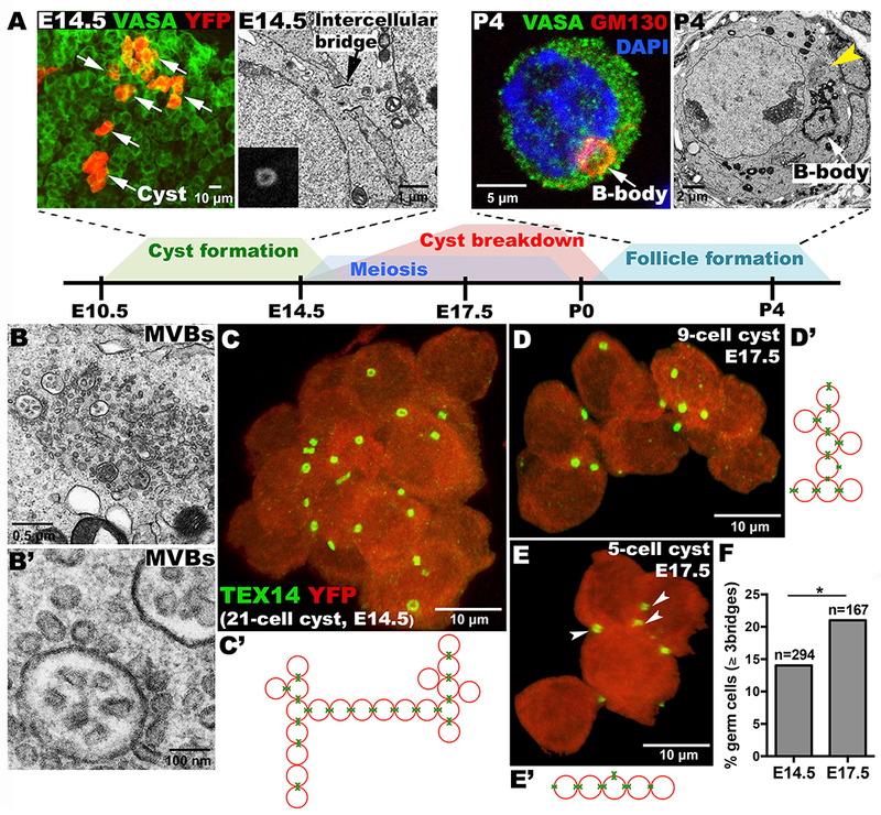
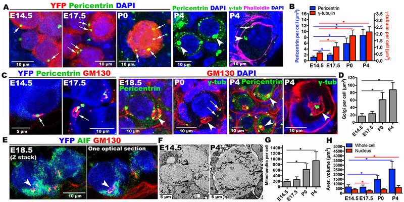
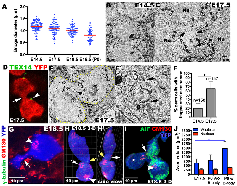
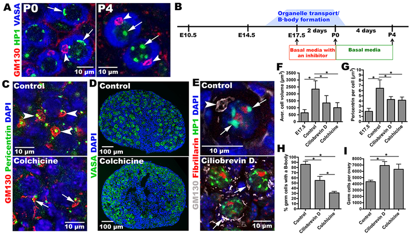
Oocytes differentiate in diverse species by receiving organelles and cytoplasm from sister germ cells while joined in germline cysts or syncytia. Mouse primordial germ cells form germline cysts, but the role of cysts in oogenesis is unknown. We find that mouse germ cells receive organelles from neighboring cyst cells and build a Balbiani body to become oocytes, whereas nurselike germ cells die. Organelle movement, Balbiani body formation, and oocyte fate determination are selectively blocked by low levels of microtubule-dependent transport inhibitors. Membrane breakdown within the cyst and an apoptosis-like process are associated with organelle transfer into the oocyte, events reminiscent of nurse cell dumping in Drosophila We propose that cytoplasmic and organelle transport plays an evolutionarily conserved and functionally important role in mammalian oocyte differentiation.


Figures

Figure 1.
Figure 1.. Germline cysts and mouse oocyte differentiation.
(A) Timeline of cyst formation and breakdown to form primordial follicles. Above, YFP-labeled single-germ-cell clone showing 6 cysts (arrows); EM showing an E14.5 ring canal (arrow), inset (Tex14-labeled ring canal); GM130-labeled Balbiani body (B-body); EM of a P4 oocyte showing B-body and multi-vesicular bodies (MVBs, yellow arrowhead). (B, B’) EM of oocyte MVBs at higher magnifications. Lineage-labeled E14.5 (C) and E17.5 (D, E) cysts showing ring canals (Tex14:green); and connection structure (C’-E’), arrowheads: 3-ring-canal cell. (F) “Branched” germ cells increase between E14.5-E17.5 (p<0.05;chi-squared).
Figure 2.
Figure 2.. Organelle enrichment and Balbiani body formation during mouse oocyte differentiation.
(A) Pericentrin or γ-tubulin-labeled centrosomes (green, arrows) increase in certain cyst cells (red) during E14.5-P4. Each P4 oocyte acquires a large Pericentrin-rich mass (arrowheads). γ-tubulin staining shows 5-6 elements (arrowhead, arrow) within a P4 germ cell delimited by phalloidin staining. (B) Histogram summarizing centrosome accumulation. (C) Golgi elements (red, arrows) often associated with centrosomes (green) also increase and generate the B-body (arrowheads). Pericentrin or γ-tubulin-labeled centrosomes (green) mostly cluster inside by P4. (D) Histogram of Golgi element content per cell. (E) Mitochondria are more abundant in the cyst cell containing a nascent B-body (arrow). (F) EM shows increases in mitochondria (arrows) between E14.5 and P4, as quantitated (G). (H) Germ cell volume increases with time. YFP=lineage marker, AIF (apoptosis-inducing factor)=mitochondria. *p<0.05. Note: GM130 antibody non-specifically stains basement membrane at E18.5-P4.
Figure 3.
Figure 3.. Balbiani body formation and oocyte differentiation are facilitated by membrane fragmentation.
(A) Ring canal (bridge) diameter decreases in size. Cyst germ cells are distinct at E14.5 (B), but frequently contain membrane gaps by E17.5 (C, arrows; F). (D, E, E’) Ring canals (D, arrowhead; E, arrows) detach from intercellular junctions (D, E’ arrow). Golgi material from a contracting cell (G, arrow) and centrosomes (H, H’, yellow arrows) move through membrane gaps into B-body-containing cells (arrowhead). (I) Cell (*) lacking most cytoplasm is connected (arrow) to mitochondria-enriched cells (green), including one with a B-body (arrowhead). (J) P0 cells containing a B-body are larger than E17.5 cells. *p<0.05.
Figure 4.
Figure 4.. Microtubule-dependent transport is important for Balbiani body formation and oocyte differentiation.
(A) Heterochromatin (arrows) re-organizes between P0 and P4. B-bodies (arrowheads). (B) Experimental design for oocyte differentiation in vitro. E17.5 ovaries are cultured with or without inhibitor for 2 d, and then cultured 4 d without inhibitor. Control ovaries efficiently develop primordial follicles (C), containing full-sized oocytes with normal B-bodies (arrowheads); ovaries treated with inhibitors form smaller cells (F) with fewer B-bodies (H) and reduced Pericentrin content (G). (D) Inhibitor-treated cultures contain more germ cells (I). (E) Heterochromatin re-organizes in controls (arrows) but remains perinuclear in inhibitor-treated cultures. *p<0.05.

Comment in

  • DEVELOPMENT. Nursing the oocyte.
    Pepling ME. Pepling ME. Science. 2016 Apr 1;352(6281):35-6. doi: 10.1126/science.aaf4943. Science. 2016. PMID: 27034359 No abstract available.
  • Nursing the Follicles.
    Chao JT, Niwa M. Chao JT, et al. Dev Cell. 2016 Apr 4;37(1):7-8. doi: 10.1016/j.devcel.2016.03.019. Dev Cell. 2016. PMID: 27046826

Similar articles

Cited by

References

    1. Sun Y-C. et al. J. Genet. Genomics 41, 87–95 (2014). - PubMed
    1. Büning J, The Insect Ovary. (New York: Chapman & Hall; ) 400pp. (1994).
    1. Matova N, Cooley L, Dev. Biol 231, 291–320 (2001). - PubMed
    1. Cox R, Spradling AC, Development 130, 1579–1590 (2003). - PubMed
    1. Jenkins VK, Timmons AK, McCall K, Trends Cell Biol. 23, 567–574 (2013). - PMC - PubMed

Publication types