. 2016 Apr;170(4):1999-2023.
doi: 10.1104/pp.15.01777. Epub 2016 Feb 25.

Composition, Assembly, and Trafficking of a Wheat Xylan Synthase Complex

Affiliations

Composition, Assembly, and Trafficking of a Wheat Xylan Synthase Complex

Nan Jiang et al. Plant Physiol. 2016 Apr.

Abstract

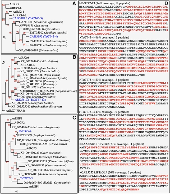
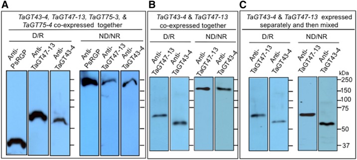
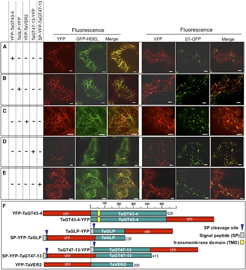
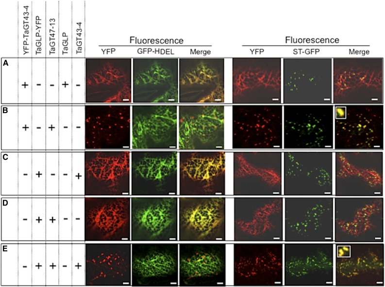
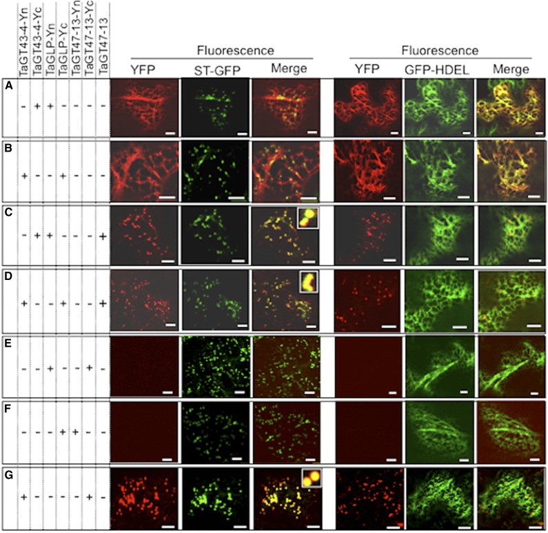
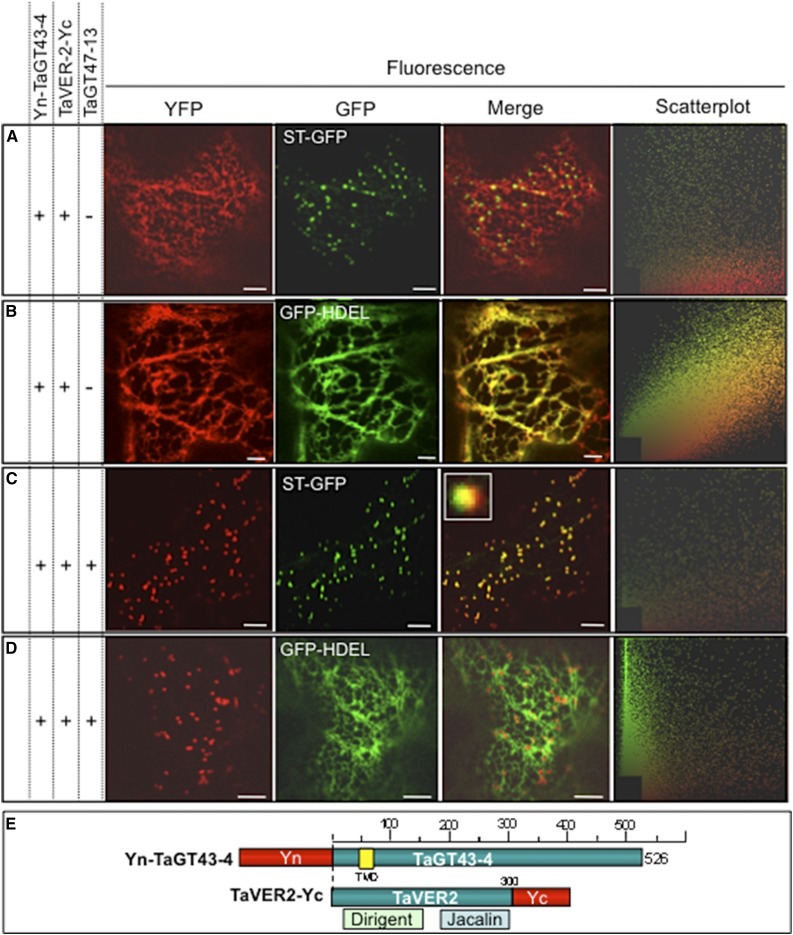
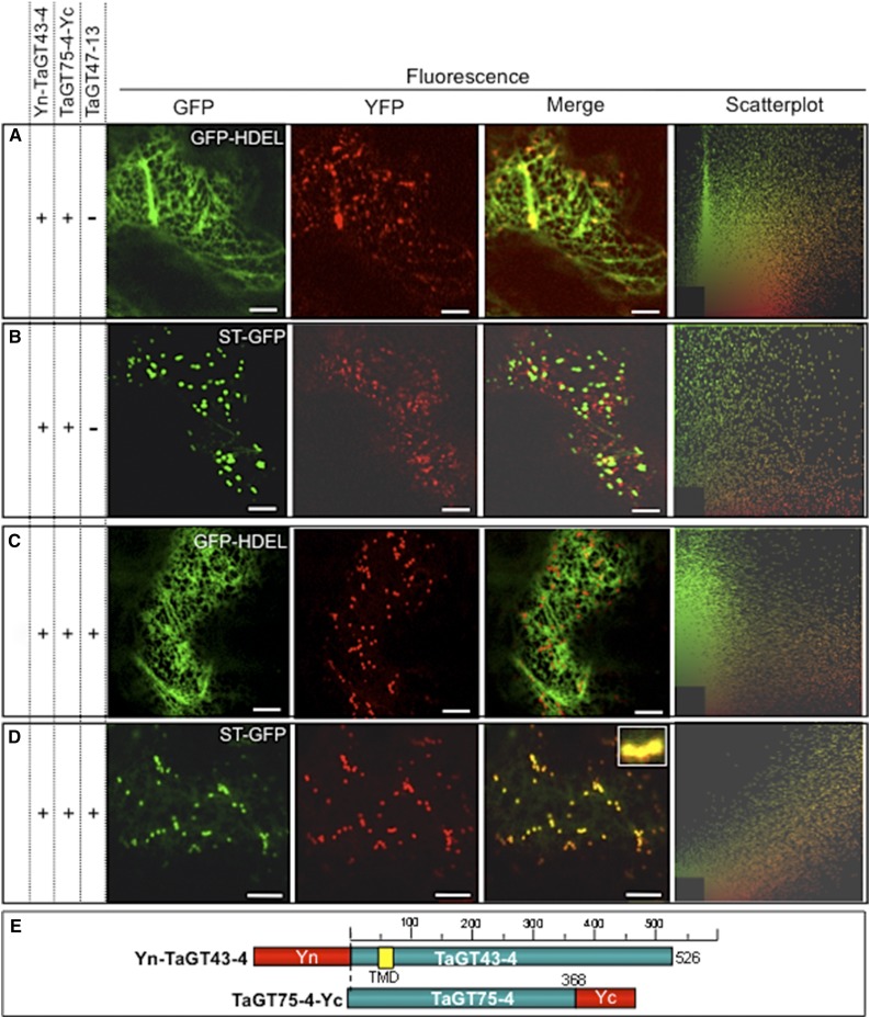
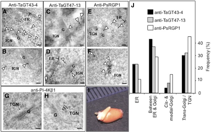
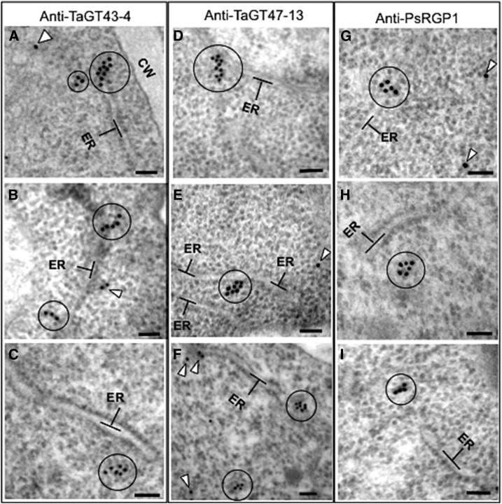
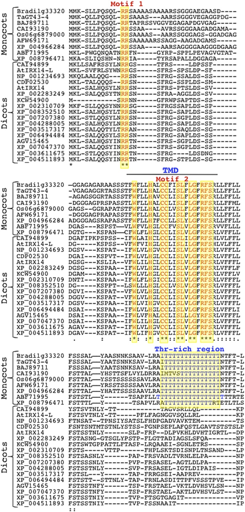
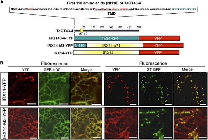
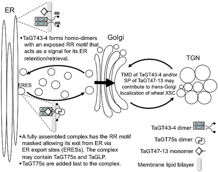
Xylans play an important role in plant cell wall integrity and have many industrial applications. Characterization of xylan synthase (XS) complexes responsible for the synthesis of these polymers is currently lacking. We recently purified XS activity from etiolated wheat (Triticum aestivum) seedlings. To further characterize this purified activity, we analyzed its protein composition and assembly. Proteomic analysis identified six main proteins: two glycosyltransferases (GTs) TaGT43-4 and TaGT47-13; two putative mutases (TaGT75-3 and TaGT75-4) and two non-GTs; a germin-like protein (TaGLP); and a vernalization related protein (TaVER2). Coexpression of TaGT43-4, TaGT47-13, TaGT75-3, and TaGT75-4 in Pichia pastoris confirmed that these proteins form a complex. Confocal microscopy showed that all these proteins interact in the endoplasmic reticulum (ER) but the complexes accumulate in Golgi, and TaGT43-4 acts as a scaffold protein that holds the other proteins. Furthermore, ER export of the complexes is dependent of the interaction between TaGT43-4 and TaGT47-13. Immunogold electron microscopy data support the conclusion that complex assembly occurs at specific areas of the ER before export to the Golgi. A di-Arg motif and a long sequence motif within the transmembrane domains were found conserved at the NH2-terminal ends of TaGT43-4 and homologous proteins from diverse taxa. These conserved motifs may control the forward trafficking of the complexes and their accumulation in the Golgi. Our findings indicate that xylan synthesis in grasses may involve a new regulatory mechanism linking complex assembly with forward trafficking and provide new insights that advance our understanding of xylan biosynthesis and regulation in plants.


Figures

Figure 1.
Figure 1.
Maximum likelihood phylogenetic analysis of proteins from GT47 (A), GT43 (B), and GT75 (C) families that were identified by proteomics (Table I). Wheat (Ta, in blue), Arabidopsis (At), and rice (Os) proteins are included for comparison. Trees were generated through the Phylogeny.fr platform using MUSCLE 3.7 (Edgar 2004) and PhyML 3.0 (Guindon et al., 2010) programs. Bootstrap values (based on 500 replicate) are indicated at the tree nodes. The scale measures evolutionary distance in substitution per amino acids. D, List of wheat proteins matched by the highest number of peptides and having the highest protein scores. Text in red indicates the amino acid sequences covered by the peptides. Protein coverage and the number of peptides matching each protein are indicated.
Figure 2.
Figure 2.
Immunoblot analysis of Triton extracts from microsomal membranes of transgenic P. pastoris cells. Triton extracts (20–25 μg proteins) were either analyzed under denaturing/reducing (D/R) conditions (1-D SDS-PAGE) or under nonreducing/nondenaturing (ND/NR) conditions (gels contain 0.05% cholate instead of SDS and no DTT) without boiling, as described by Zeng et al. (2010). A, Triton extracts from yeast cells coexpressing TaGT43-4, TaGT47-13, TaGT75-3, and TaGT75-4. B, Triton extracts from yeast cells coexpressing only TaGT43-4 and TaGT47-13. C, Triton extracts from yeast cells expressing TaGT43-4 or TaGT47-13 individually that were mixed (1:1 ratio) before analysis. Polyacrylamide gels were analyzed by immunoblot using anti-TaGT43-4, anti-TaGT47-13, and anti-PsRGP1 antibodies. Molecular weight markers (kD) are indicated.
Figure 3.
Figure 3.
Subcellular localization of YFP-tagged TaGT43-4, TaGT47-13, TaGLP, and TaVER2 in epidermal cells of tobacco leaves. ER marker, GFP-HDEL, and α(2,6)sialyltransferase fused to GFP (ST-GFP), trans-Golgi marker are used to show the colocalization with YFP-tagged proteins. GFP and YFP fluorescence are shown in green and red, respectively, and their colocalization appears in yellow. Tagged proteins were transiently expressed individually (indicated by a “+” sign on the left of the figure). YFP-TaGT43-4, TaGLP-YFP, and YFP-TaVER2 localize to the ER (A–C, respectively). While TaGT47-13-YFP localizes with ST-GFP (D), SP-YFP-TaGT47-13 localizes with GFP-HDEL (E). Bars = 10 μm. Schematic presentations of YFP-tagged constructs used in this study are shown in F.
Figure 4.
Figure 4.
Trafficking of YFP-TaGT43-4 and TaGLP-YFP when transiently expressed in combination with untagged TaGT47-13, TaGT43-4, and/or TaGLP in epidermal cells of tobacco leaves. Coinfiltrated constructs are indicated by the “+” at the left of the figure. ER marker (GFP-HDEL) and trans-Golgi marker (ST-GFP) were used to show the colocalization with YFP-tagged proteins. GFP and YFP fluorescence are shown in green and red, respectively, and their colocalization appears in yellow. Note that YFP-TaGT43-4 export from the ER relies on the presence of TaGT47-13 (B), while the presence of TaGLP does not affect its localization (A). TaGLP-YFP export from the ER requires the presence of both TaGT43-4 and TaGT47-13 (E), while the presence of TaGT43-4 alone does not affect its localization (C). The insets in merge pictures in B and E show the overlap between YFP and ST-GFP. Bars = 10 μm.
Figure 5.
Figure 5.
TaGT43-4, TaGT47-13, and TaGLP assemble in the ER before export to the trans-Golgi. Protein-protein interactions were visualized via BiFC (split-YFP). Coinfiltrated constructs are indicated by the “+” at the left of the figure. ER marker (GFP-HDEL) and trans-Golgi marker (ST-GFP) were included to show the localization of the reconstituted YFP. GFP and YFP fluorescence are shown in green and red, respectively, and their colocalization (merge) appears in yellow. The insets in merge pictures in C, D, and G show the overlap between YFP and ST-GFP. Bars = 10 μm
Figure 6.
Figure 6.
TaGT43-4, TaVER2, and TaGT47-13 assemble in the ER before export to the Golgi. Protein-protein interactions were investigated using BiFC (split-YFP). The data show that TaGT43-4 interacts with TaVER2 in the ER to form a complex that is retained in the ER (A and B) until interaction with untagged TaGT47-13, which results in the export of the complex from the ER to a Golgi compartment that has partial overlap with trans-Golgi (C and D). ST-GFP and GFP-HDEL, and trans-Golgi and ER markers were used to show the colocalization of the assembled YFP. GFP and YFP fluorescence are shown in green and red, respectively, and their colocalization (merge) appears in yellow. Two-dimensional scatterplots on the right display the degree of overlap between the red and green in the images. The inset in merge picture in C shows the limited overlap between YFP and ST-GFP. Bars = 10 μm. Schematic presentations of YFP-tagged constructs used in this study are shown in E. TaVER2 is a soluble protein containing two domains: a dirigent domain at the NH2-terminal end and a lectin domain (Jacalin) at the COOH-terminal end.
Figure 7.
Figure 7.
TaGT43-4 interacts with TaGT75-4 at specific areas of the ER (A and B). The inclusion of untagged TaGT47-13 results in ER export of “TaGT43-4/TaGT75-4” complex to trans-Golgi (C and D). Protein-protein interaction between TaGT43-4 and TaGT75-4 was investigated using BiFC (split-YFP). ST-GFP and GFP-HDEL, and trans-Golgi and ER markers were used to show the colocalization of the assembled YFP. GFP and YFP fluorescence are shown in green and red, respectively, and their colocalization (merge) appears in yellow. Two-dimensional scatterplots on the right display the degree of overlap between the red and green in the images. The inset in merge picture in D shows the overlap between YFP and ST-GFP. Bars = 10 μm. Schematic presentations of YFP-tagged constructs used in this experiment are shown in E.
Figure 8.
Figure 8.
Self-assembly of TaGT43-4, TaGT75-3, TaGT75-4, TaGT47-13, and TaGLP was investigated through BiFC (split-YFP). Both TaGT43-4 and TaGLP can form homodimers that localize to the ER (A and D, respectively). TaGT75-3 and TaGT75-4 can form homodimers mostly localize to the Golgi (G and I, respectively) and can also form heterodimers (H). TaGT47-13 cannot self-assemble (F). Interaction of TaGT43-4 homodimers with TaGT47-13 results in the shift of the fluorescence to the trans-Golgi (B), while TaGLP homodimers are retained in the ER until interaction with both TaGT43-4 and TaGT47-13 (E). ER marker (GFP-HDEL) and trans-Golgi marker (ST-GFP) were included to show the localization of the assembled YFP. GFP and YFP fluorescence are shown in green and red, respectively, and their colocalization (merge) appears in yellow. Bars = 10 μm.
Figure 9.
Figure 9.
Immunoelectron micrographs of anti-TaGT43-4 (A and B), anti-TaGT47-13 (C and D), anti-PsRGP1 (E and F), and anti-PI-4Kβ1 (G and H) in shoots of 3-d-old etiolated wheat seedlings (I). Particles are marked with arrowheads. Individual immunogold particles (marked with arrowhead) are seen at the ER surface, between the ER and the cis-Golgi, and on the trans-Golgi and TGN. Note that anti-PsRGP1 labels on ER membranes were exclusively associated with the clusters (see Fig. 10), as no individual labels on ER membranes were observed outside of these clusters. Frequency (the number of particles counted from ∼50 cells) of immunogold labeling localization is presented in J. Bars = 100 nm.
Figure 10.
Figure 10.
Immunoelectron micrographs of anti-TaGT43-4 (A–C), anti-TaGT47-13 (DF), and anti-PsRGP1 (G–I) in shoots of 3-d-old etiolated wheat seedlings. Circles indicate clusters of 3 to 10 immunogold particles seen as protrusions/extensions from the ER (A, B, D, and G) or as clusters detached but near the ER (C, F, H, and I). Arrowheads indicate individual immunogold particles. Bars = 100 nm.
Figure 11.
Figure 11.
Alignment of the NH2-terminal regions (first 100 amino acids) of TaGT43-4 and 25 homologous proteins of the GT43 family. Sequences were obtained from NCBI, Phytozome 10.3, or CAZy database. The alignment was generated through MUSCLE 3.7 program in the Phylogeny.fr platform. The two conserved motifs (RR and Wx3Hx2CCx2Sx2LGxRFS in red) as well as a Thr-rich region specific to monocot proteins (in blue) are highlighted in yellow. Monocot and dicot proteins with their accession numbers are indicated.
Figure 12.
Figure 12.
Confocal images of epidermal cells of tobacco leaves transiently expressing Arabidopsis IRX14-YFP (B) or IRX14-M3-YFP, which encodes a protein having the first 71 amino acids replaced with the first 110 amino acids from TaGT43-4 (Nt110; C). Schematic presentations of these constructs are shown in A. ST-GFP and GFP-HDEL, and trans-Golgi and ER markers were used to show YFP localization. GFP and YFP fluorescence are shown in green and red, respectively, and their colocalization (merge) appears in yellow. NH2-terminal region of TaGT43-4 is sufficient to retain IRX14-M3-YFP in the ER. Bars = 10 μm.
Figure 13.
Figure 13.
Model for the assembly and trafficking of wheat XSC. This model assumes that the central core of wheat XSC is formed of TaGT43-4 and TaGT47-13 but can include TaGT75s, TaGLP, and/or TaVER2. The assembly of this core complex occurs in the ER through a coupled folding and binding mechanism and starts with TaGT43-4 forming homodimers. However, the trafficking of the functionally assembled complex is signal mediated and involves a di-Arg (RR) motif at the NH2-terminal region of TaGT43-4. This RR motif is exposed in unassembled TaGT43-4 homodimers but is masked in a functional TaGT43-4/TaGT47-13 complex. Unmasked RR motif acts as an ER retention/retrieval signal, while masked RR motif allows the functional complex to escape this retention/retrieval process and exit the ER from ERESs. The accumulation of the complex in trans-Golgi is hypothesized to be under the control of the TMD of TaGT43-4 and the NH2-terminal secretion SP of TaGT47-13, acting as a cryptic TMD. This model assumes that TaGT75s are added to the complex at the ERESs before export to the Golgi.

References

    1. Anders N, Wilkinson MD, Lovegrove A, Freeman J, Tryfona T, Pellny TK, Weimar T, Mortimer JC, Stott K, Baker JM, et al. (2012) Glycosyl transferases in family 61 mediate arabinofuranosyl transfer onto xylan in grasses. Proc Natl Acad Sci USA 109: 989–993 - PMC - PubMed
    1. Aoki D, Lee N, Yamaguchi N, Dubois C, Fukuda MN (1992) Golgi retention of a trans-Golgi membrane protein, galactosyltransferase, requires cysteine and histidine residues within the membrane-anchoring domain. Proc Natl Acad Sci USA 89: 4319–4323 - PMC - PubMed
    1. Atmodjo MA, Sakuragi Y, Zhu X, Burrell AJ, Mohanty SS, Atwood JA III, Orlando R, Scheller HV, Mohnen D (2011) Galacturonosyltransferase (GAUT)1 and GAUT7 are the core of a plant cell wall pectin biosynthetic homogalacturonan:galacturonosyltransferase complex. Proc Natl Acad Sci USA 108: 20225–20230 - PMC - PubMed
    1. Bar-Peled M, Raikhel NV (1997) Characterization of AtSEC12 and AtSAR1. Proteins likely involved in endoplasmic reticulum and Golgi transport. Plant Physiol 114: 315–324 - PMC - PubMed
    1. Barlowe C. (2003) Signals for COPII-dependent export from the ER: what’s the ticket out? Trends Cell Biol 13: 295–300 - PubMed

Publication types

MeSH terms

LinkOut - more resources