

Large Epiphrenic Diverticula: A Rare Case Presentation
- PMID: 26918097
- PMCID: PMC4745591
- DOI: 10.4081/cp.2015.784
Large Epiphrenic Diverticula: A Rare Case Presentation
Abstract
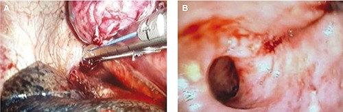
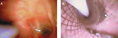
A 70-year old female was admitted to hospital with heartburn and chronic halitosis since 5 years. She was on proton pump inhibitors for the same. Her complaints worsened during the last one-year. Workup comprising of esophago-gastro-duodenoscopy, esophageal manometry, 3D computed tomography scan showed right-sided epiphrenic diverticula measuring 10x10 cm with wide mouth about 5 cm with hypertensive lower esophageal sphincter. Patient underwent a video assisted thoracoscopic surgery for esophageal diverticulectomy using two 45 mm staplers. On day 5, the patient developed leak, which was managed by a covered esophageal stent placement. Patient started on oral feeds from day 3 and the esophageal leak healed completely within 2 weeks. Literature suggests that esophageal leaks treated conservatively took approximately 30-40 days on an average for healing. Literature search did not reveal esophageal leak managed by stent with faster recovery (2 weeks). This is one of the largest epiphrenic diverticuli reported in literature.
Keywords: Epiphrenic diverticulum; achalasia; leak; stent.
Conflict of interest statement
Conflict of interest: the authors declare no potential conflict of interest.
Figures


References
-
- Palanivelu C, Rangarajan M, Maheshkumaar GS, Senthilkumar R. Minimally invasive surgery combined with preoperative endoscopy for symptomatic middle and lower esophageal diverticula: a single institute’s experience. Surg Laparosc Endosc Percutan Tech 2008;18:133-8. - PubMed
-
- Guerra JM, Zuil M, Garcia I, Moreno E. Epiphrenic diverticula, esophageal carcinoma and esophagopleural fistula. Hepato-Gastroenterol 2001;48:718-9. - PubMed
-
- Costantini M, Zaninotto G, Rizzetto C, et al. Oesophageal diverticula. Best Pract Res Clin Gastroenterol 2004;18:3-17. - PubMed
-
- Skinner DB, Belsey RH. Esophageal spasm and diverticulum (Management of esophageal disease). Philadelphia, PA: W.B. Saunders; 1988. pp 31-452.
-
- Benacci JC, Deschamps C, Trastek VF, et al. Epiphrenic diverticulum: results of surgical treatment. Ann Thorac Surg 1993;55:1109-13. - PubMed
Publication types
LinkOut - more resources
Full Text Sources
Other Literature Sources