Review
. 2015 Dec 16:4:F1000 Faculty Rev-1460.
doi: 10.12688/f1000research.7006.1. eCollection 2015.

Drug-Induced Metabolic Acidosis

Affiliations
Review

Drug-Induced Metabolic Acidosis

Amy Quynh Trang Pham et al. F1000Res. .

Abstract

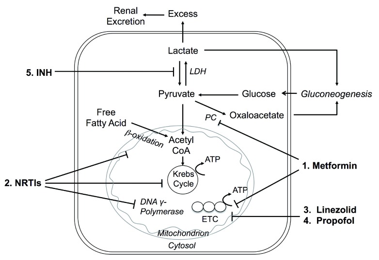
Metabolic acidosis could emerge from diseases disrupting acid-base equilibrium or from drugs that induce similar derangements. Occurrences are usually accompanied by comorbid conditions of drug-induced metabolic acidosis, and clinical outcomes may range from mild to fatal. It is imperative that clinicians not only are fully aware of the list of drugs that may lead to metabolic acidosis but also understand the underlying pathogenic mechanisms. In this review, we categorized drug-induced metabolic acidosis in terms of pathophysiological mechanisms, as well as individual drugs' characteristics.

Keywords: MALA; acidosis; drug-induced; metabolic.


Conflict of interest statement

Competing interests: The authors have no competing interests.

No competing interests were disclosed.

Figures

Figure 1.
Figure 1.. Excretion of acid and ways to jeopardize the system.
1. A strong non-volatile acid HA dissociates to release H + and poses an immediate threat to plasma pH. 2. Bicarbonate buffers the H + and generates CO 2, which is expelled in the lungs and results in depletion of body HCO 3 -. Non-bicarbonate buffers (collectively referred to as B) carry the H + until the kidneys excrete it. 3. The kidneys split CO 2 into H + and HCO 3 - and selectively secrete H + into the lumen and HCO 3 - into the blood. In addition, any excess H + from the body fluid is also excreted. 4. Most H + excreted in the urine is carried by urinary buffers (UBs). 5. Some organic anions (A) (e.g. lactate, ketoanions) can be metabolized to regenerate the HCO 3 -. If A is not metabolizable (e.g. phosphate or sulfate), it is excreted in the urine. * Two possible ways by which metabolic acidosis can occur.
Figure 2.
Figure 2.. Mechanisms of drug-induced metabolic acidosis.
1. Increased exogenous ingestion of acidic precursors that are converted into strong acids. 2. Loss of alkali from kidney or GI tract. 3. Increased endogenous production of strong organic acids. 4. Compromised renal net acid excretion by inhibition of the renin-angiotensin-aldosterone system (RAAS), impaired proximal tubule (PT), or distal tubule (DT) H + secretion.
Figure 3.
Figure 3.. Mechanisms of drug-induced lactic acidosis.
1. Metformin inhibits pyruvate carboxylase (PC) → inhibits hepatic gluconeogenesis → excess lactate . Metformin also inhibits complex I of the mitochondrial electron transport chain (ETC) → increases NADH/NAD + ratio → blocks the entry of pyruvate into the tricarboxylic acid (TCA) cycle . LDH = lactate dehydrogenase 2. In vitro, nucleoside reverse transcriptase inhibitors (NRTIs) inhibit β-oxidation, the tricarboxylic acid (Krebs) cycle, and DNA γ-polymerase → mitochondrial dysfunction and loss of transcription of essential enzymes → hepatic steatosis (increased triglycerides), myopathy, pancreatitis, nephrotoxicity, and lactic acidosis . 3. Linezolid may cross-react with mammalian cellular processes → disrupts mitochondrial protein synthesis involved in ETC , . 4. Propofol may inhibit coenzyme Q and cytochrome C at Complex IV in ETC, and also inhibit mitochondrial fatty acid metabolism . 5. Isoniazid inhibits metabolism of lactate to pyruvate .
Figure 4.
Figure 4.. Mechanisms of drug-induced distal H + secretion.
1. Cyclooxygenase (COX) inhibitors and β-blockers interfere with release of renin, leading to hyperkalemia with metabolic acidosis , . 2. Angiotensin-converting enzyme inhibitors (ACEIs), aldosterone receptor blockers (ARBs), and renin inhibitors all interfere with the renin-angiotensin-aldosterone system (RAAS), causing hyperkalemia with hyperchloremic metabolic acidosis . 3. Heparin and ketoconazole , interfere with aldosterone synthesis. 4. Spironolactone and eplerenone block aldosterone receptors , . 5. Na + channel blockers lead to reduced net negative charge in lumen in cortical collecting ducts (CCD), which reduces K + and H + excretion and causes hyperkalemia and acidosis , – . 6. Calcineurin inhibitors interfere with Na, K-ATPase in the principal cell decreasing transepithelial K secretion and H + secretion, cause vasoconstriction of afferent glomerular arterioles, and decrease glomerular filtration rate and alter filtration fraction , . 7. Lithium causes a voltage-dependent defect for H + secretion and decreases H +-ATPase activity . 8. Amphotericin B binds to sterol in mammalian cell membranes , forming intramembranous pores which increase permeability and back diffusion of H +.
Figure 5.
Figure 5.. Mechanisms of proximal tubule (PT) and drug-induced Fanconi syndrome.
1. CA inhibitors cause bicarbonaturia and hyperchloremic metabolic acidosis in the elderly and patients with renal failure and diabetes . 2. Antineoplastic platinum-containing agents , and DNA-alkylating agents damage proximal tubule cells through accumulation and induced cell apoptosis. 3. Anti-viral/HIV drugs , – , valproic acid (VPA) , and outdated tetracycline interfere with mitochondrial function within proximal tubule cells, leading to tubular dysfunction. 4. Aminoglycosides , , induce acidosis with unclear mechanisms . 5. Deferasirox increases hemodynamic iron removal, causes vacuolization of proximal tubular epithelial cells , and elevates iron absorption in various organs. All could lead to acidosis.

References

    1. Moe OW, Fuster D, Alpern RJ: Common acid-base disorders. In: Goldman L, Wachter RM, Hollander H, editors. Hosp Med 2nd ed. Philadelphia: Lippincott, William & Wilkins;2005;1055–65.
    1. Wiederkehr MR, Moe OW: Treatment of metabolic acidosis. In: Massry SG, Suki WK, editors. Therapy of Renal Diseases and Related Disorders 4th ed: Springer; In press,2011.
    1. Moe OW, Fuster D: Clinical acid-base pathophysiology: disorders of plasma anion gap. Best Pract Res Clin Endocrinol Metab. 2003;17(4):559–74. 10.1016/S1521-690X(03)00054-X - DOI - PubMed
    1. McMartin KE, Ambre JJ, Tephly TR: Methanol poisoning in human subjects. Role for formic acid accumulation in the metabolic acidosis. Am J Med. 1980;68(3):414–8. 10.1016/0002-9343(80)90113-8 - DOI - PubMed
    1. Jacobsen D, Hewlett TP, Webb R, et al. : Ethylene glycol intoxication: evaluation of kinetics and crystalluria. Am J Med. 1988;84(1):145–52. 10.1016/0002-9343(88)90024-1 - DOI - PubMed