Review
. 2015 Dec 7:4:F1000 Faculty Rev-1408.
doi: 10.12688/f1000research.6623.1. eCollection 2015.

Capturing relevant extracellular matrices for investigating cell migration

Affiliations
Review

Capturing relevant extracellular matrices for investigating cell migration

Patricia Keely et al. F1000Res. .

Abstract

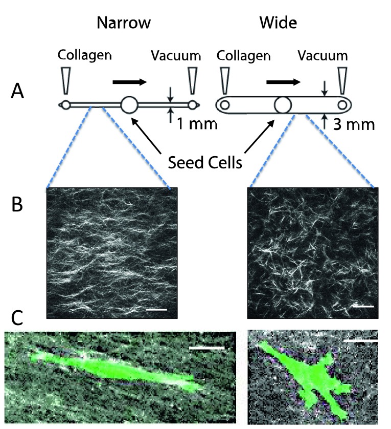
Much progress in understanding cell migration has been determined by using classic two-dimensional (2D) tissue culture platforms. However, increasingly, it is appreciated that certain properties of cell migration in vivo are not represented by strictly 2D assays. There is much interest in creating relevant three-dimensional (3D) culture environments and engineered platforms to better represent features of the extracellular matrix and stromal microenvironment that are not captured in 2D platforms. Important to this goal is a solid understanding of the features of the extracellular matrix-composition, stiffness, topography, and alignment-in different tissues and disease states and the development of means to capture these features.

Keywords: ECM; Matrix remodeling; cell migration; extracellular matrix; stromal microenvironment.


Conflict of interest statement

Competing interests: The authors declare that they have no competing interests.

No competing interests were disclosed.

Figures

Figure 1.
Figure 1.. Collagen alignment around tumors facilitates invasion.
( A) Second harmonic generation (SHG) image of a normal mouse mammary gland. Collagen appears white. ( B) SHG of a mouse mammary PyMT tumor. Note the straightened and aligned collagen perpendicular to the tumor boundary. Both images are reprinted with permission from Provenzano et al. (2008). ( C) Intravital image of human MDA-MB-231 breast carcinoma cells in mouse mammary gland. Cells are transfected with green fluorescent protein. Collagen appears white. Note that cells polarize along collagen. ( D) Zoom of boxed region in ( C). The arrow points to a single collagen fiber in the field of view. Scale bars = 50 μm.
Figure 2.
Figure 2.. Use of fibronectin-coated fibers on a suspended platform.
The platform was created by using the spinneret-based tunable engineered parameter (STEP) technique as described by Nain et al. (2009). ( A) STEP manufacturing platform and scanning electron microscope images of fibers fabricated (i) same diameter crosshatch, (ii) mix of diameters in three layers, and (iii) varying orientations. ( B) Immunofluorescence images of single cells in (i) spindle, (ii) parallel with star showing leading edge, and (iii) polygonal shapes (red: f-actin stress fibers, blue: nucleus, and green: paxillin focal adhesion clusters). Scale bars: 10 µm (Ai and Aii), 20 µm (Aiii), 50 µm ( B).
Figure 3.
Figure 3.. Creation of aligned and random collagen gels in microfluidic channels.
( A) Design of microchannels. Collagen is neutralized and pre-incubated for several minutes prior to pulling it across a narrow (1 mm wide × 250 μm tall) or wide (3 mm wide × 250 μm tall) microfluidic chamber. The chambers are incubated for several hours at 37°C, and then the mask for the central port is removed, allowing cells to be seeded into the central port. Cells are incubated and imaged by time-lapse microscopy for 3–4 days. ( B) Second harmonic generation image of collagen in a narrow and wide chamber demonstrates aligned fibers in the narrow chamber. ( C) Cells in aligned matrices (narrow channel) exhibit elongated morphology and minimal protrusions, whereas cells in random matrices (wide channel) have more abundant protrusions. MDA-MB-231 breast carcinoma cells were transfected with LifeAct-GFP. All images are reprinted with permission from Riching et al. .

References

    1. Tscharntke M, Pofahl R, Chrostek-Grashoff A, et al. : Impaired epidermal wound healing in vivo upon inhibition or deletion of Rac1. J Cell Sci. 2007;120(Pt 8):1480–90. 10.1242/jcs.03426 - DOI - PubMed
    2. F1000 Recommendation

    1. Alexander S, Weigelin B, Winkler F, et al. : Preclinical intravital microscopy of the tumour-stroma interface: invasion, metastasis, and therapy response. Curr Opin Cell Biol. 2013;25(5):659–71. 10.1016/j.ceb.2013.07.001 - DOI - PubMed
    1. Petrie RJ, Gavara N, Chadwick RS, et al. : Nonpolarized signaling reveals two distinct modes of 3D cell migration. J Cell Biol. 2012;197(3):439–55. 10.1083/jcb.201201124 - DOI - PMC - PubMed
    2. F1000 Recommendation

    1. Tozluoğlu M, Tournier AL, Jenkins RP, et al. : Matrix geometry determines optimal cancer cell migration strategy and modulates response to interventions. Nat Cell Biol. 2013;15(7):751–62. 10.1038/ncb2775 - DOI - PubMed
    1. Friedl P, Sahai E, Weiss S, et al. : New dimensions in cell migration. Nat Rev Mol Cell Biol. 2012;13(11):743–7. 10.1038/nrm3459 - DOI - PubMed