. 2016:1:15001.
doi: 10.1038/sigtrans.2015.1. Epub 2016 Jan 28.

Human IgG1 antibodies suppress angiogenesis in a target-independent manner

Affiliations

Human IgG1 antibodies suppress angiogenesis in a target-independent manner

Sasha Bogdanovich et al. Signal Transduct Target Ther. 2016.

Abstract

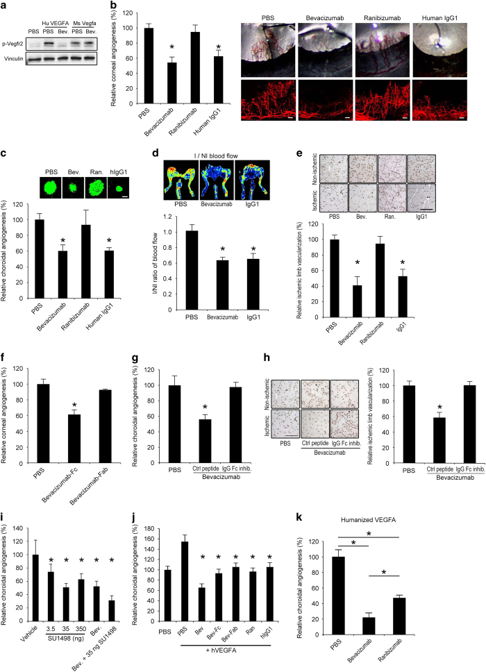
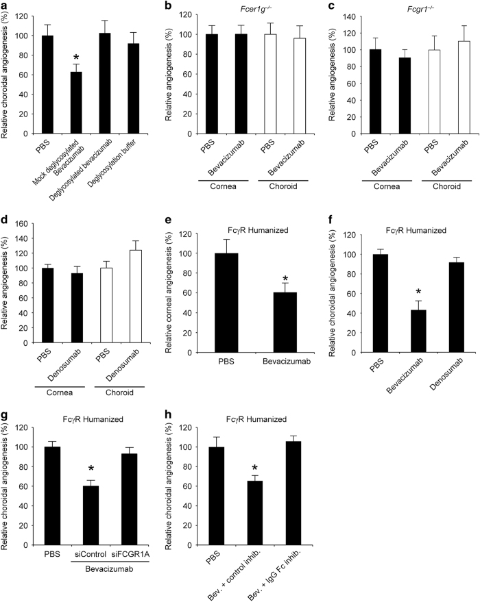
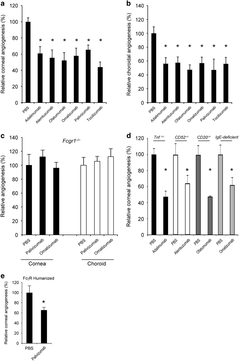
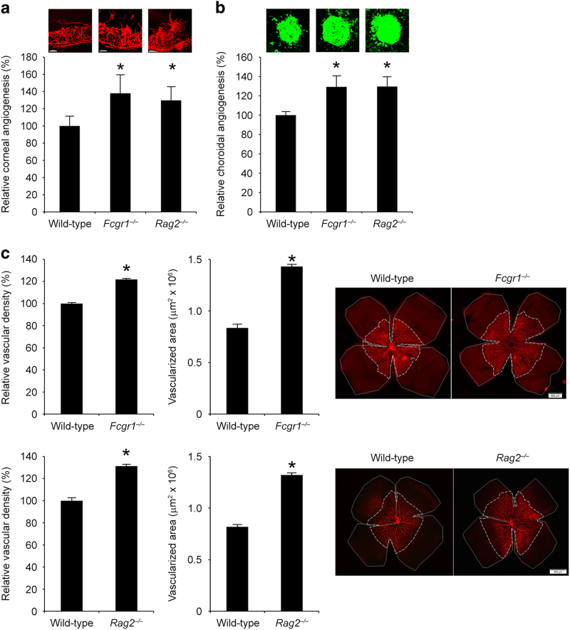
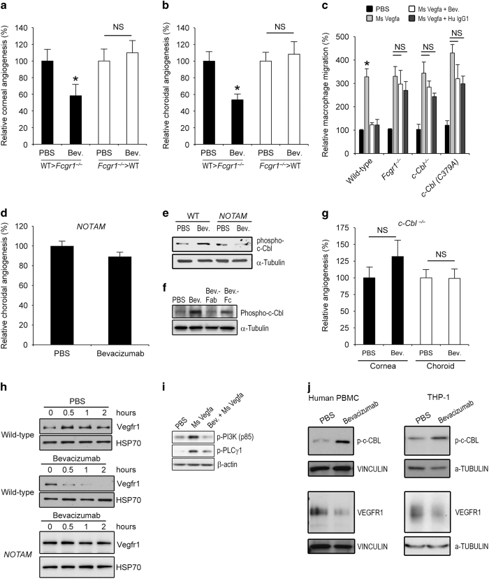
Aberrant angiogenesis is implicated in diseases affecting nearly 10% of the world's population. The most widely used anti-angiogenic drug is bevacizumab, a humanized IgG1 monoclonal antibody that targets human VEGFA. Although bevacizumab does not recognize mouse Vegfa, it inhibits angiogenesis in mice. Here we show bevacizumab suppressed angiogenesis in three mouse models not via Vegfa blockade but rather Fc-mediated signaling through FcγRI (CD64) and c-Cbl, impairing macrophage migration. Other approved humanized or human IgG1 antibodies without mouse targets (adalimumab, alemtuzumab, ofatumumab, omalizumab, palivizumab and tocilizumab), mouse IgG2a, and overexpression of human IgG1-Fc or mouse IgG2a-Fc, also inhibited angiogenesis in wild-type and FcγR humanized mice. This anti-angiogenic effect was abolished by Fcgr1 ablation or knockdown, Fc cleavage, IgG-Fc inhibition, disruption of Fc-FcγR interaction, or elimination of FcRγ-initated signaling. Furthermore, bevacizumab's Fc region potentiated its anti-angiogenic activity in humanized VEGFA mice. Finally, mice deficient in FcγRI exhibited increased developmental and pathological angiogenesis. These findings reveal an unexpected anti-angiogenic function for FcγRI and a potentially concerning off-target effect of hIgG1 therapies.


Conflict of interest statement

JA is a co-founder of iVeena Holdings, iVeena Pharmaceuticals, iVeena Delivery Systems and Inflammasome Therapeutics, and has received honoraria from Allergan and research funding from Olix Pharmaceuticals unrelated to this work. JA and SDF are named as inventors on patent applications filed by the University of Kentucky relating to the technology described in this work. MRC is listed as an inventor on patents covering alemtuzumab and MRC and KLA are listed as inventors on patents covering the Fc mutated forms of alemtuzumab.

Figures

Figure 1
Figure 1
Bevacizumab inhibited mouse angiogenesis via Fc region. (a) Western blot shows that bevacizumab inhibited Vegfr2 phosphorylation (pVegfr2) in Py4 mouse hemangioma endothelial cells when treated with human VEGFA, but not when treated with mouse Vegfa, after 10 min. Image representative of three experiments. (b) Bevacizumab and human IgG1, but not ranibizumab, decreased corneal angiogenesis in wild-type mice. Area of angiogenesis was measured 10 days after suture injury and normalized to PBS group. n=10–38. Representative photos of wild-type mouse eyes (upper row) and corneal flat mounts (lower row) showing reduced growth of blood vessels (CD31+, red) in eyes treated with bevacizumab or human IgG1, but not in eyes treated with ranibizumab. Scale bars, 100 μm. (c) Bevacizumab and human IgG1, but not ranibizumab, suppressed choroidal angiogenesis in wild-type mice 7 days after laser injury compared with PBS (experiment performed in JA laboratory). Images depict representative choroidal angiogenesis lesions (endothelial cells stained in green) in each treatment group. n=12–20. (d, e) Treatment of ischemic hind limb with bevacizumab or human IgG1 in wild-type mice suppressed muscle revascularization and decreased blood vessel perfusion, as seen in representative laser Doppler perfusion images (top), and measured blood flow in the ischemic limbs (bottom), normalized to the contralateral non-ischemic limbs, 7 days after surgery. n=6. I/NI, ischemic/non-ischemic. Bevacizumab and human IgG1, but not ranibizumab, treatment of ischemic limbs reduced muscle angiogenesis (CD31+, brown) as seen in representative images of muscle CD31 immunolocalization (e), and quantification of muscle CD31 immunolocalization (bottom), normalized to the contralateral non-ischemic limbs. (f) The Fc fragments, not the Fab fragment, of bevacizumab suppressed corneal angiogenesis in wild-type mice. Area of angiogenesis was measured 10 days after suture injury and normalized to PBS group. n=10–38. (g) Co-administration of a peptide that prevents IgG-Fc binding to FcγR, but not a control peptide, blocked inhibition of choroidal angiogenesis by bevacizumab in wild-type mice. (h) Co-administration of an IgG-Fc inhibitory peptide, but not a control peptide, blocked inhibition of muscle angiogenesis (CD31+, brown) by bevacizumab, as seen in representative images of muscle CD31 immunolocalization (left), and quantification of muscle CD31 immunolocalization (right), normalized to the contralateral non-ischemic limbs. Scale bar, 100 μm. n=6. (i) Bevacizumab suppressed choroidal angiogenesis in wild-type mice to the same extent as SU1498, a small molecule tyrosine kinase inhibitor of Vegfr2. Combined administration of bevacizumab and SU1498 suppressed choroidal angiogenesis to a greater extent than either of the agents alone. n=6. (j) Choroidal angiogenesis, augmented by administration of human VEGFA, was suppressed to similar extents by ranibizumab, bevacizumab-Fab, bevacizumab-Fc and human IgG1; and, to a greater extent, by bevacizumab. n=6–8. (k) Bevacizumab suppressed choroidal angiogenesis to a greater extent than ranibizumab in the humanized VEGFA mouse, a transgenic model that expresses a VEGFA protein that can be neutralized by both bevacizumab and ranibizumab. n=6. Results are means±s.e.m. *P<0.05 compared with PBS (bh, k) or with vehicle (i) or with PBS+human VEGFA (j).
Figure 2
Figure 2
Bevacizumab inhibited mouse angiogenesis via FcγRI. (a) Deglycosylated bevacizumab did not suppress choroidal angiogenesis in wild-type mice; however, choroidal angiogenesis was inhibited by bevacizumab subjected to mock treatment. The deglycosylation buffer had no effect on choroidal angiogenesis. n=8–14. (b) Bevacizumab did not suppress corneal or choroidal angiogenesis in Fcer1g−/− mice. n=8–10. (c) Bevacizumab did not inhibit corneal or choroidal angiogenesis in Fcgr1−/− mice. No significant difference between groups. n=10–13. (d) Denosumab did not suppress corneal or choroidal angiogenesis in wild-type mice. n=6–8. No significant difference between groups. (e) Bevacizumab inhibited corneal angiogenesis in FcγR humanized mice. n=8. Results are means±s.e.m. *P<0.05 compared with PBS. (f) Bevacizumab, but not denosumab, inhibited choroidal angiogenesis in FcγR humanized mice. n=6–8. (g) Co-administration of a 17+2-nt cholesterol conjugated human FCGR1A siRNA, but not a 17+2-nt cholesterol-conjugated control Luc siRNA, blocked inhibition of choroidal angiogenesis by bevacizumab in FcγR humanized mice. n=8. (h) Co-administration of an IgG-Fc inhibitory peptide, but not a control peptide, blocked inhibition of choroidal angiogenesis by bevacizumab in FcγR humanized mice. n=8. Results are means±s.e.m. *P<0.05 compared with PBS (a, eh).
Figure 3
Figure 3
Bevacizumab interacted with, induced phosphorylation of, and upregulated abundance of FcγRI in vivo. (a) Wild-type mouse corneas that had been administered biotinylated bevacizumab or biotinylated denosumab following suture injury were subjected to streptavidin pull-down and immunoblotting for mouse FcγRI. Biotinylated bevacizumab, but not denosumab, interacted with mouse FcγRI in vivo. Anti-streptavidin immunoblotting confirmed efficient pull-down of both biotinylated antibodies. (b) FcγR humanized mouse corneas that had been administered bevacizumab or PBS following suture injury were subjected to immunoprecipitation of human FcγRI followed by immunoblotting for human IgG1 or phosphotyrosine. Bevacizumab, but not PBS, interacted with and induced phosphorylation of human FcγRI in vivo. Reprobing confirmed efficient immunoprecipitation of human FcγRI in both bevacizumab- and PBS-treated corneas. Each image is representative of three experiments (a, b). (c) Bevacizumab, but not PBS, increased Fcgr1 mRNA abundance in RAW264.7 mouse macrophages and in wild-type mouse corneas following suture injury, as monitored by real-time reverse transcription PCR, and FcγRI protein abundance in RAW264.7 cells, as monitored by western blotting. Densitometry of FcγRI normalized to Vinculin shown. n=4–6. Results are means±s.e.m. *P<0.05 compared with PBS.
Figure 4
Figure 4
Human IgG1s inhibited mouse angiogenesis via FcγRI. Treatment with the human IgG1 antibodies adalimumab, alemtuzumab, ofatumumab, omalizumab, palivizumab or tocilizumab reduced (a) corneal and (b) choroidal angiogenesis in wild-type mice. n=8–19. (c) Palivizumab and Omalizumab did not inhibit corneal or choroidal angiogenesis in Fcgr1−/− mice. n=6–8. No significant difference between groups. (d) Adalimumab, a human anti-TNFα monoclonal antibody, inhibited corneal angiogenesis in Tnf−/− mice. n=9. Alemtuzumab, a humanized anti-CD52 monoclonal antibody, inhibited corneal angiogenesis in CD52−/− mice. n=8. Ofatumumab, a human anti-CD20 monoclonal antibody, inhibited corneal angiogenesis in CD20−/− mice. n=8. Omalizumab, a humanized anti-IgE monoclonal antibody, inhibited corneal angiogenesis in IgE-deficient mice. n=10. (e) Palivizumab inhibited choroidal angiogenesis in FcγR humanized mice. Results are means±s.e.m. *P<0.05 compared with PBS (a, b, d, e).
Figure 5
Figure 5
Endogenous Igs suppressed mouse angiogenesis. Corneal angiogenesis area (a) and choroidal angiogenesis volume (b) are greater in Fcgr1−/− and Rag2−/− mice compared with wild-type mice. n=8–20. (c) The vascular density and total area of vascularized retina at postnatal day 4 is greater in Fcgr1−/− and Rag2−/− mice compared with wild-type mice. n=8. Results are means±s.e.m. *P<0.05 compared to wild-type mice (ac). Vascular density in the retina is normalized to wild-type mice. Representative flat mounts of corneal (a, red), choroidal (b, green) and retinal (c, red), vessels are shown.
Figure 6
Figure 6
Bevacizumab inhibited angiogenesis via macrophage FcγRI and c-Cbl. Bevacizumab suppressed corneal (a) and choroidal (b) angiogenesis in Fcgr1−/− mice transplanted with wild-type mouse bone marrow, but not in wild-type mice receiving Fcgr1−/− bone marrow. n=11–16. (c) Bevacizumab and human IgG1 inhibited mouse Vegfa-induced migration, over 12 h, of bone marrow-derived macrophages isolated from wild-type mice but not from Fcgr1−/−, c-Cbl−/− or c-Cbl (C379A) mice, which lack E3 ubiquitin ligase activity. n=3. Results are means±s.e.m. *P<0.05 compared with PBS (ac). (d) Bevacizumab did not inhibit choroidal angiogenesis in NOTAM mice. n=10. (e) Western blot shows induction of c-Cbl phosphorylation in wild-type mouse BMDMs treated with bevacizumab for 15 min. Protein loading was assessed by α-Tubulin abundance. (f) Western blot shows in vivo induction of c-Cbl phosphorylation in wild-type mouse corneas following suture injury that were treated with bevacizumab or its Fc fragment, but not by its Fab fragment. (g) Bevacizumab did not inhibit corneal or choroidal angiogenesis in c-Cbl−/− mice. n=11–30. NS, no significant difference between groups. (h) Western blots show time-dependent Vegfr1 degradation in wild-type but not NOTAM mouse BMDMs treated with bevacizumab. Protein loading was assessed by HSP70 abundance. (i) Western blots show that RAW264.7 mouse macrophages pre-treated with bevacizumab, but not PBS, 2 h before stimulation with mouse Vegfa, exhibited reduced phosphorylation of PI3K and PLCγ1 at 10 min after Vegfa exposure. Protein loading was assessed by β-actin abundance. (j) Western blots show induction of c-Cbl phosphorylation in human peripheral blood mononuclear cells (PBMC) or in THP-1 human monocytic cells, treated with bevacizumab, but not PBS, for 15 min. Treatment with bevacizumab, but not PBS, reduced VEGFR1 abundance in human PBMCs and THP-1 cells. Protein loading was assessed by Vinculin or α-Tubulin abundance. Images representative of three experiments (e, f, hj).

References

    1. Nelson AL , Dhimolea E , Reichert JM . Development trends for human monoclonal antibody therapeutics. Nat Rev Drug Discov 2010; 9: 767–774. - PubMed
    1. Presta LG , Chen H , O'Connor SJ , Chisholm V , Meng YG , Krummen L et al. Humanization of an anti-vascular endothelial growth factor monoclonal antibody for the therapy of solid tumors and other disorders. Cancer Res 1997; 57: 4593–4599. - PubMed
    1. Ferrara N , Hillan KJ , Gerber HP , Novotny W . Discovery and development of bevacizumab, an anti-VEGF antibody for treating cancer. Nat Rev Drug Discov 2004; 3: 391–400. - PubMed
    1. Group CR , Martin DF , Maguire MG , Ying GS , Grunwald JE , Fine SL et al. Ranibizumab and bevacizumab for neovascular age-related macular degeneration. N Engl J Med 2011; 364: 1897–1908. - PMC - PubMed
    1. Liang WC , Wu X , Peale FV , Lee CV , Meng YG , Gutierrez J et al. Cross-species vascular endothelial growth factor (VEGF)-blocking antibodies completely inhibit the growth of human tumor xenografts and measure the contribution of stromal VEGF. J Biol Chem 2006; 281: 951–961. - PubMed

Publication types