Review
. 2016 Oct 1;594(19):5471-90.
doi: 10.1113/JP271764. Epub 2016 May 12.

Diverse roles for ionotropic glutamate receptors on inhibitory interneurons in developing and adult brain

Affiliations
Review

Diverse roles for ionotropic glutamate receptors on inhibitory interneurons in developing and adult brain

Gülcan Akgül et al. J Physiol. .

Abstract

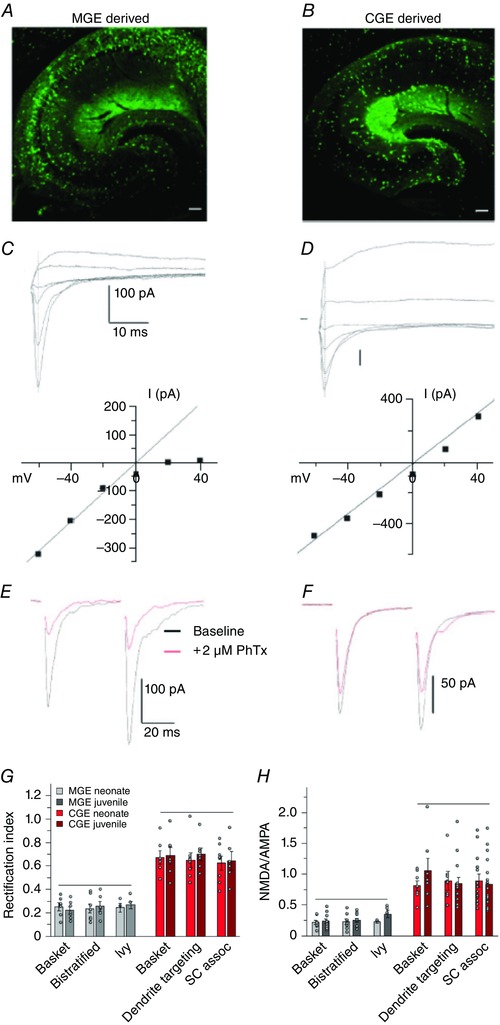
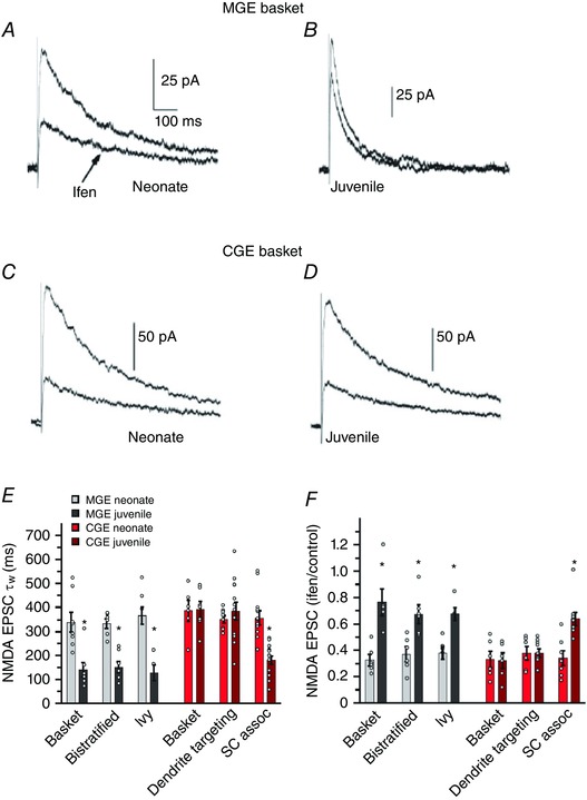
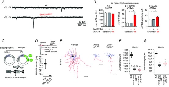
Glutamate receptor-mediated recruitment of GABAergic inhibitory interneurons is a critical determinant of network processing. Early studies observed that many, but not all, interneuron glutamatergic synapses contain AMPA receptors that are GluA2-subunit lacking and Ca(2+) permeable, making them distinct from AMPA receptors at most principal cell synapses. Subsequent studies demonstrated considerable alignment of synaptic AMPA and NMDA receptor subunit composition within specific subtypes of interneurons, suggesting that both receptor expression profiles are developmentally and functionally linked. Indeed glutamate receptor expression profiles are largely predicted by the embryonic origins of cortical interneurons within the medial and caudal ganglionic eminences of the developing telencephalon. Distinct complements of AMPA and NMDA receptors within different interneuron subpopulations contribute to the differential recruitment of functionally divergent interneuron subtypes by common afferent inputs for appropriate feed-forward and feedback inhibitory drive and network entrainment. In contrast, the lesser-studied kainate receptors, which are often present at both pre- and postsynaptic sites, appear to follow an independent developmental expression profile. Loss of specific ionotropic glutamate receptor (iGluR) subunits during interneuron development has dramatic consequences for both cellular and network function, often precipitating circuit inhibition-excitation imbalances and in some cases lethality. Here we briefly review recent findings highlighting the roles of iGluRs in interneuron development.


Figures

Figure 1
Figure 1. AMPARs on interneuron subtypes show embryonic origin‐specific properties
The vast majority of hippocampal interneurons originate from two distinct embryonic regions, the MGE and CGE. MGE‐ (A) and CGE‐ (B) derived cohorts of inhibitory interneurons are shown here labelled with eGFP expression driven by Nkx2.1 or 5ht3ra promoter activity, respectively. C and D, glutamatergic synapses onto MGE‐ and CGE‐derived interneurons differ in their synaptic properties. Traces are representative of Schaffer collateral‐evoked total glutamate receptor (AMPAR and NMDAR)‐mediated EPSCs recorded in voltage clamp configuration from MGE‐derived (C) and CGE‐derived (D) interneurons of the CA1 stratum radiatum, at holding potentials ranging from −60 mV to +40 mV. The vertical line that crosses the raw traces indicates the time point for the extraction of the AMPAR‐mediated fast component of the EPSCs. The plot of these data reveals an inwardly rectifying current–voltage relationship for AMPARs at synapses onto cells derived from the MGE (C) and an essentially linear current–voltage relationship for AMPARs onto CGE‐derived interneurons (D). At positive holding potentials Schaffer collateral–MGE interneuron EPSCs exhibit only a relatively small fast NMDAR component indicating that the contribution of NMDARs at these synapses is low (C and H). In contrast NMDAR‐mediated EPSCs at Schaffer collateral–CGE synapses are relatively large and slow (D and H). E and F, AMPARs on MGE‐ and CGE‐derived interneurons show differential sensitivity to block by extracellular polyamines. The traces show AMPAR‐mediated EPSCs before (black) and after (red) application of philanthotoxin (PhTx) for representative recordings from MGE‐derived (E) and CGE‐derived (F) interneurons. PhTx blocks AMPAR‐mediated currents in a use‐dependent manner in MGE‐derived interneurons to a greater extent compared to CGE‐derived interneurons verifying the Ca2+‐permeable properties of AMPARs at this synapse. G and H, graphs summarizing a detailed developmental analysis of the NMDAR‐ and AMPAR‐mediated EPSCs. The current–voltage relationship remains unchanged across two developmental time points (postnatal day P6–9 and P17–21) indicating that the AMPAR subunit composition is not susceptible to developmental regulation (G). Across a similar developmental age range NMDA/AMPA receptor ratios are low (∼1:5) in three morphologically distinct subtypes of MGE‐derived interneurons in CA1 and ∼1 in three distinct CGE‐derived interneuron subtypes. Reproduced from Matta et al. (2013) with permission from the authors.
Figure 2
Figure 2. NMDAR subunit expression in MGE‐derived hippocampal interneurons show developmental regulation
A–D, channel gating of NMDARs accelerates and loses GluN2B antagonist (ifenprodil) sensitivity in MGE‐derived interneurons whereas NMDARs in CGE‐derived interneurons have properties that persist through development. The traces are representative evoked NMDAR‐mediated EPSCs recorded at +40 mV holding potential from MGE‐derived basket cells (A and B) and CGE‐derived basket cells (C and D) from a neonate (A and C) and a juvenile (B and D) upon Schaffer collateral stimulation in the absence (upper traces) or presence (lower traces) of ifenprodil. In both MGE‐ and CGE‐derived basket cells of neonate (A and C), the peak amplitude of the current is markedly reduced indicating GluN2B‐rich NMDARs in these interneurons. Similar antagonist application is unable to reduce evoked current in MGE‐derived basket cell of juvenile indicating a significant loss of GluN2B subunits from NMDAR pool in this synapse. Juvenile NMDAR currents also possess faster kinetics than neonate consistent with a GluN2B–GluN2A switch (B). In contrast ifenprodil block is significant in CGE‐derived basket cell across both developmental time points indicating that NMDA receptor subunit expression does not undergo developmental regulation in CGE‐derived interneurons (D). E, graph summarizes the detailed analysis of the NMDAR EPSC decay kinetics weighted time constant (τw) for both neonate and juvenile MGE‐ and CGE‐derived interneurons at two different developmental time points, neonate (P6–9) and juvenile (P17–21). F, graph summarizes the detailed analysis of the developmental regulation of ifenprodil sensitivity expressed as the ratio of the NMDAR EPSC peak amplitude measured in the presence of ifenprodil divided by the control NMDA EPSC peak amplitude. Reproduced from Matta et al. (2013) with permission from the authors.
Figure 3
Figure 3. Genetic deletion of NMDARs reveals control of both synaptic and anatomical properties
A and B, genetic deletion of the GluN2B subunit of NMDARs results in abnormal maturation of glutamatergic synapses in hippocampal interneurons. In the transgenic line the gene coding for GluN2B is flanked with loxP cassettes and deleted upon breeding with GAD67:Cre mice. A, representative traces of spontaneous EPSCs from wild‐type (wt; upper trace) and GluN2B knockout (GluN2BΔGAD67; lower trace) fast spiking GAD67‐positive interneurons in stratum oriens. In GluN2B knockout interneurons, there is an apparent reduction in spontaneous events, as well as large amplitude events indicating that the number of active synapses has decreased while more AMPARs are incorporated into those remaining. B, graphs show the group data for analysis of the firing properties of the wild‐type and knockout interneurons (left), interspike intervals of spontaneous (s)EPSCs as representation of event frequency (IEI, middle), and mean amplitude of events (right). C–G, loss of either all NMDARs or only the GluN2B subunit alone in cortical interneurons results in abnormal anatomy. C, the genetic strategy to create the conditional knockout animals. Both transgenic animal and virus technology were used to knock out all or the GluN2B‐rich subgroup of NMDARs in CGE‐derived cortical interneurons. The genes coding for GluN1 or GluN2B were deleted in Dlx5/6‐expressing interneurons of targeted knockin mice in which the genes coding for GluN1 or GluN2B are flanked with loxP cassettes. Upon electroporation of viruses that carry the Cre‐expressing sequence downstream of Dlx5/6 promoter, glun1 or glun2b gene deletions occur. (E, embryonic day.) D, the traces are representative evoked EPSCs at −70 mV (inward current) and +40 mV (outward current, black trace and no current, red trace) holding potentials show the loss of NMDAR‐mediated currents (red trace) in GluN1‐knockout interneurons. The graph below shows the group data. E, reconstructions of reelin‐positive interneurons of wild‐type (left), GluN1‐ (middle) and GluN2B‐ (right) knockout mice. Both GluN1 and GluN2B loss result in shrinkage of dendrites and axons in RE‐positive cortical interneurons and GluN2B loss is enough to create this anatomical abnormality. Scale bars, 50 μm. Axons are shown in red, dendrites in blue. The data are summarized with the graphs for axonal (F) and dendritic (G) length analysis of the RE‐positive interneuron morphology. A and B are reproduced with permission from Kelsch et al. (2014). C–G are reproduced with permission from De Marco Garcia et al. (2015).

References

    1. Akbarian S, Sucher NJ, Bradley D, Tafazzoli A, Trinh D, Hetrick WP, Potkin SG, Sandman CA, Bunney WE Jr & Jones EG (1996). Selective alterations in gene expression for NMDA receptor subunits in prefrontal cortex of schizophrenics. J Neurosci 16, 19–30. - PMC - PubMed
    1. Akgul G & Wollmuth LP (2013). Synapse‐associated protein 97 regulates the membrane properties of fast‐spiking parvalbumin interneurons in the visual cortex. J Neurosci 33, 12739–12750. - PMC - PubMed
    1. Ali AB, Deuchars J, Pawelzik H & Thomson AM (1998). CA1 pyramidal to basket and bistratified cell EPSPs: dual intracellular recordings in rat hippocampal slices. J Physiol 507, 201–217. - PMC - PubMed
    1. Ali AB & Thomson AM (1998). Facilitating pyramid to horizontal oriens‐alveus interneurone inputs: dual intracellular recordings in slices of rat hippocampus. J Physiol 507, 185–199. - PMC - PubMed
    1. Allen K & Monyer H (2015). Interneuron control of hippocampal oscillations. Curr Opin Neurobiol 31, 81–87. - PubMed

Publication types

MeSH terms

Substances

LinkOut - more resources