

Severe chronic cerebral hypoperfusion induces microglial dysfunction leading to memory loss in APPswe/PS1 mice
- PMID: 26918610
- PMCID: PMC4914254
- DOI: 10.18632/oncotarget.7689
Severe chronic cerebral hypoperfusion induces microglial dysfunction leading to memory loss in APPswe/PS1 mice
Abstract
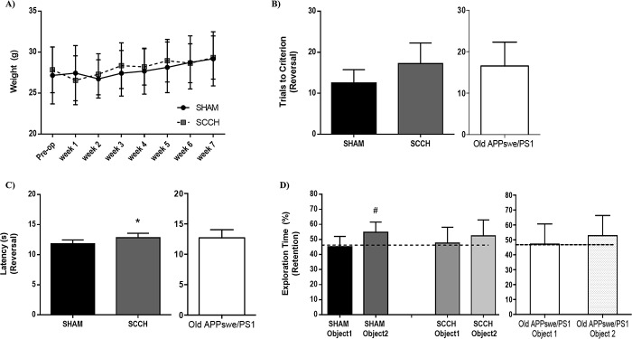
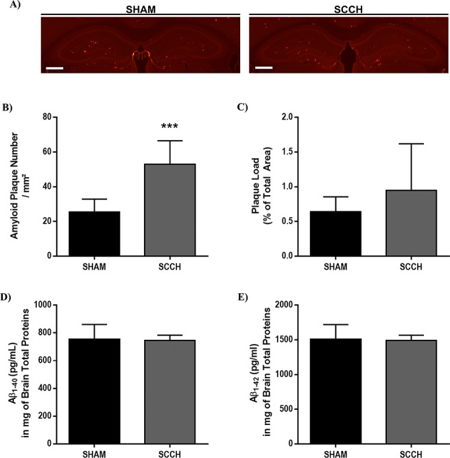
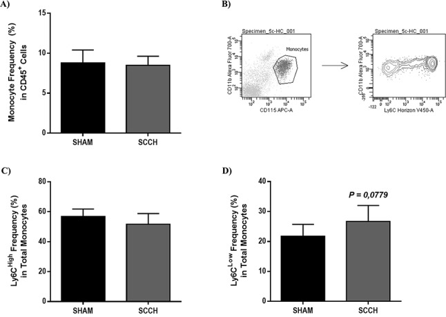
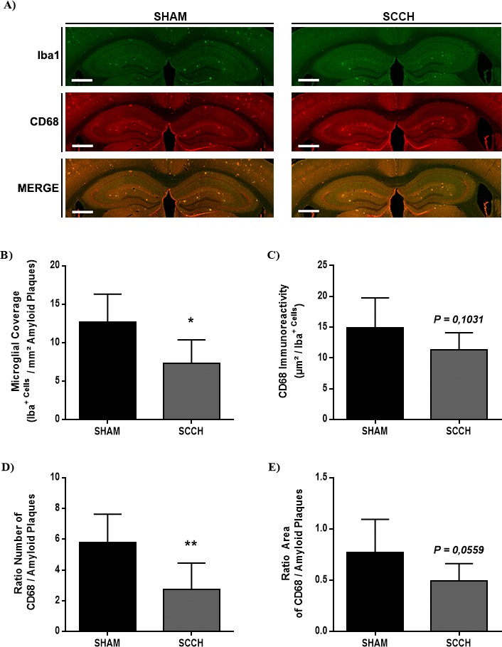
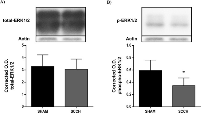
Cerebral vasculature plays a key role in controlling brain homeostasis. Cerebral vasculature dysfunction, associated to irregularities in cerebral blood perfusion, has been proposed to directly contribute to Alzheimer's disease (AD) pathogenesis. More precisely, chronic cerebral hypoperfusion, which impairs brain homeostasis, was demonstrated to take place even before cognitive decline. However, the mechanisms underlying the implication of chronic cerebral hypoperfusion in AD pathogenesis remain elusive. Therefore, this study aims at investigating the role of severe chronic cerebral hypoperfusion (SCCH) in AD pathogenesis. For this purpose, SCCH was induced in young APPswe/PS1 in order to evaluate the progression of AD-like pathology in these mice. We observed that SCCH accelerated the cognitive decline of young APPswe/PS1 mice, which was associated with an increased amyloid plaque number in brain parenchyma. In addition, SCCH reduced the activity of extracellular signal-regulated kinases 1/2 (ERK1/2), which has been shown to play an important role in the adaptive responses of neurons. Importantly, SCCH impaired the function of microglial cells, which are implicated in amyloid-β (Aβ) elimination. In vitro approaches underlined the ability of a low-glucose microenvironment to decrease the general activity and phagocytic capacity of microglia. By using a new model of SCCH, our study unravels new insights into the implication of severe chronic cerebral hypoperfusion in AD pathogenesis, mainly by altering microglial cell activity and consequently Aβ clearance.
Keywords: Alzheimer’s disease; Gerotarget; cerebral hypoperfusion; microglia.
Figures
Similar articles
-
Tissue-Plasminogen Activator Attenuates Alzheimer's Disease-Related Pathology Development in APPswe/PS1 Mice.Neuropsychopharmacology. 2016 Apr;41(5):1297-307. doi: 10.1038/npp.2015.279. Epub 2015 Sep 9. Neuropsychopharmacology. 2016. PMID: 26349911 Free PMC article.
-
Increased Spontaneous Central Bleeding and Cognition Impairment in APP/PS1 Mice with Poorly Controlled Diabetes Mellitus.Mol Neurobiol. 2016 May;53(4):2685-97. doi: 10.1007/s12035-015-9311-2. Epub 2015 Jul 9. Mol Neurobiol. 2016. PMID: 26156287 Free PMC article.
-
Role of Suppressor of Cytokine Signaling 3 (SOCS3) in Altering Activated Microglia Phenotype in APPswe/PS1dE9 Mice.J Alzheimers Dis. 2017;55(3):1235-1247. doi: 10.3233/JAD-160887. J Alzheimers Dis. 2017. PMID: 27814300
-
Chronic cerebral hypoperfusion: a key mechanism leading to vascular cognitive impairment and dementia. Closing the translational gap between rodent models and human vascular cognitive impairment and dementia.Clin Sci (Lond). 2017 Sep 28;131(19):2451-2468. doi: 10.1042/CS20160727. Print 2017 Oct 1. Clin Sci (Lond). 2017. PMID: 28963120 Review.
-
Cerebral hypoperfusion and glucose hypometabolism: Key pathophysiological modulators promote neurodegeneration, cognitive impairment, and Alzheimer's disease.J Neurosci Res. 2017 Apr;95(4):943-972. doi: 10.1002/jnr.23777. Epub 2016 Jun 27. J Neurosci Res. 2017. PMID: 27350397 Review.
Cited by
-
Multifocal Cerebral Microinfarcts Modulate Early Alzheimer's Disease Pathology in a Sex-Dependent Manner.Front Immunol. 2022 Jan 31;12:813536. doi: 10.3389/fimmu.2021.813536. eCollection 2021. Front Immunol. 2022. PMID: 35173711 Free PMC article.
-
Neurovascular Alterations in Vascular Dementia: Emphasis on Risk Factors.Front Aging Neurosci. 2021 Sep 10;13:727590. doi: 10.3389/fnagi.2021.727590. eCollection 2021. Front Aging Neurosci. 2021. PMID: 34566627 Free PMC article.
-
Optical coherence tomography-based assessment of retinal vascular pathology in cerebral small vessel disease.Neurol Res Pract. 2020 May 15;2:13. doi: 10.1186/s42466-020-00062-4. eCollection 2020. Neurol Res Pract. 2020. PMID: 33324919 Free PMC article.
-
Cognitive Dysfunction after Heart Disease: A Manifestation of the Heart-Brain Axis.Oxid Med Cell Longev. 2021 Aug 18;2021:4899688. doi: 10.1155/2021/4899688. eCollection 2021. Oxid Med Cell Longev. 2021. PMID: 34457113 Free PMC article. Review.
-
Pathophysiology of blood brain barrier dysfunction during chronic cerebral hypoperfusion in vascular cognitive impairment.Theranostics. 2022 Jan 16;12(4):1639-1658. doi: 10.7150/thno.68304. eCollection 2022. Theranostics. 2022. PMID: 35198062 Free PMC article. Review.
References
-
- Wimo A, Jonsson L, Winbald B. An estimate of the worldwide prevalence and direct loss costs of dementia in 2003. Dement Geriatr Cogn Disord. 2006;21:175–181. - PubMed
-
- Alzheimer A, Stelzmann RA, Schnitzlein HN, Murtagh FR. An english translation of Alzheimer's 1907 paper, ‘Uber eine eigenartige erkankaung der hirnrinde’. Clin Anat. 1995;8:429–431. - PubMed
-
- Iadecola C, Gorelick PB. Converging pathogenic mechanisms in vascular and neurodegenerative dementia. Stroke. 2003;34:335–337. - PubMed
-
- Marchesi VT. Alzheimer's dementia begins as a disease of small blood vessels, damaged by oxidative-induced inflammation and dysregulated amyloid metabolism: Implication for early detection and therapy. FASEB J. 2011;25:5–13. - PubMed
MeSH terms
Substances
LinkOut - more resources
Full Text Sources
Other Literature Sources
Medical
Molecular Biology Databases
Miscellaneous
