. 2016 Feb 26;11(2):e0149897.
doi: 10.1371/journal.pone.0149897. eCollection 2016.

Inflammation Promotes Expression of Stemness-Related Properties in HBV-Related Hepatocellular Carcinoma

Affiliations

Inflammation Promotes Expression of Stemness-Related Properties in HBV-Related Hepatocellular Carcinoma

Te-Sheng Chang et al. PLoS One. .

Erratum in

Abstract

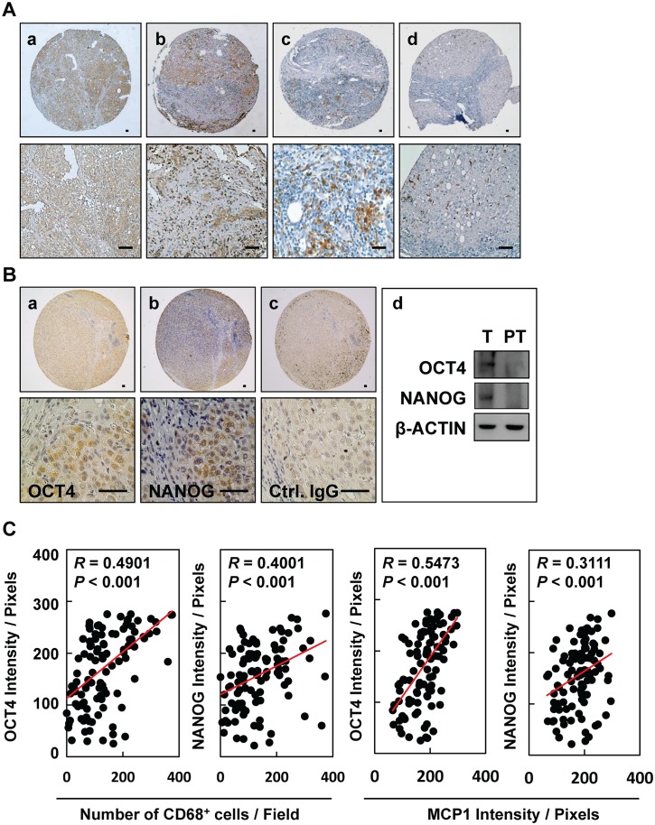
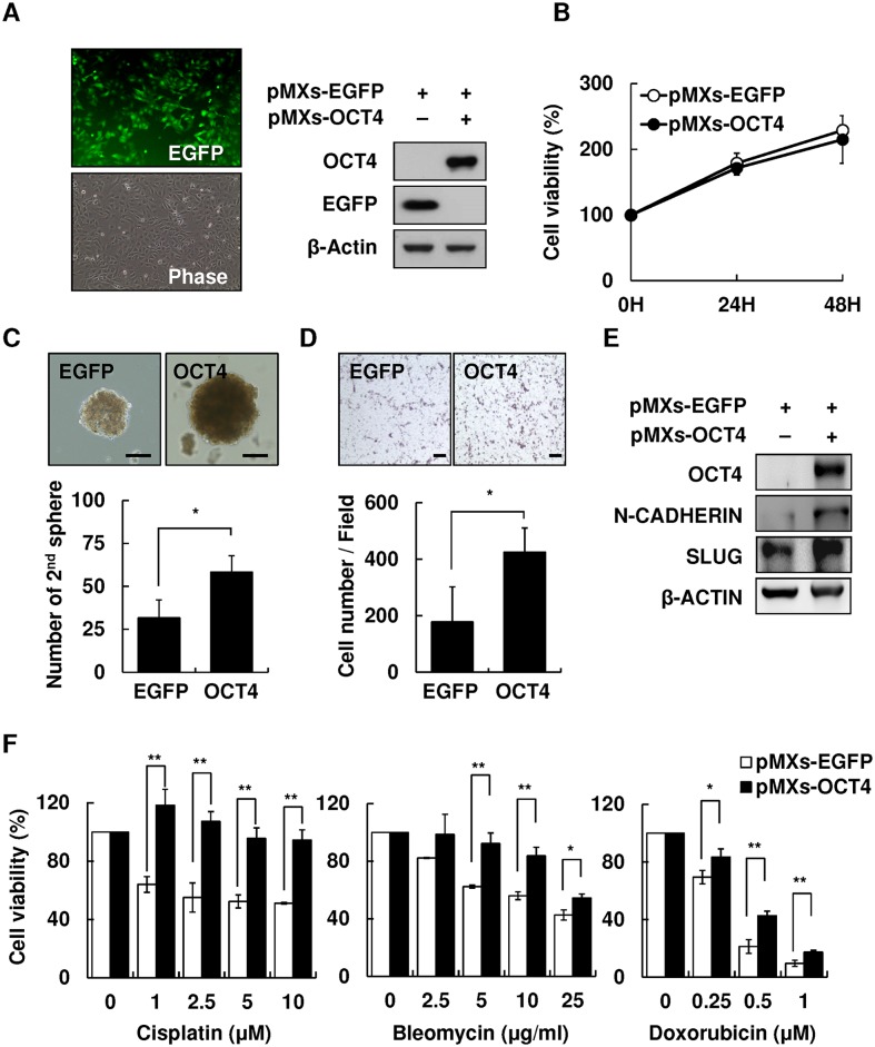
The expression of cancer stemness is believed to reduce the efficacy of current therapies against hepatocellular carcinoma (HCC). Understanding of the stemness-regulating signaling pathways incurred by a specific etiology can facilitate the development of novel targets for individualized therapy against HCC. Niche environments, such as virus-induced inflammation, may play a crucial role. However, the mechanisms linking inflammation and stemness expression in HCC remain unclear. Here we demonstrated the distinct role of inflammatory mediators in expressions of stemness-related properties involving the pluripotent octamer-binding transcription factor 4 (OCT4) in cell migration and drug resistance of hepatitis B virus-related HCC (HBV-HCC). We observed positive immunorecognition for macrophage chemoattractant protein 1 (MCP-1)/CD68 and OCT4/NANOG in HBV-HCC tissues. The inflammation-conditioned medium (inflamed-CM) generated by lipopolysaccharide-stimulated U937 human leukemia cells significantly increased the mRNA and protein levels of OCT4/NANOG preferentially in HBV-active (HBV+HBsAg+) HCC cells. The inflamed-CM also increased the side population (SP) cell percentage, green fluorescent protein (GFP)-positive cell population, and luciferase activity of OCT4 promoter-GFP/luciferase in HBV-active HCC cells. Furthermore, the inflamed-CM upregulated the expressions of insulin-like growth factor-I (IGF-I)/IGF-I receptor (IGF-IR) and activated IGF-IR/Akt signaling in HBV-HCC. The IGF-IR phosphorylation inhibitor picropodophyllin (PPP) suppressed inflamed-CM-induced OCT4 and NANOG levels in HBV+HBsAg+ Hep3B cells. Forced expression of OCT4 significantly increased the secondary sphere formation and cell migration, and reduced susceptibility of HBV-HCC cells to cisplatin, bleomycin, and doxorubicin. Taking together, our results show that niche inflammatory mediators play critical roles in inducing the expression of stemness-related properties involving IGF-IR activation, and the upregulation of OCT4 contributes to cancer migration and drug resistance of HBV-HCC cells. Findings in this paper would provide potential targets for a therapeutic strategy targeting on inflammatory environment for HBV-HCC.


Conflict of interest statement

Competing Interests: The authors have declared that no competing interests exist.

Figures

Fig 1
Fig 1. Expression of MCP-1/CD68 and pluripotent transcription factors OCT4 and NANOG in HBV-HCC tissues.
Immunohistochemical staining of (A) MCP-1 (a, b) and CD68 (c, d) and (B) OCT4 (a) and NANOG (b) in HBV-HCC tissues. (d) Expression of OCT and NANOG in tumor (T) and peritumor (PT) tissues from patients. Bar = 50 um. (C) The correlations between the inflammatory levels (CD68 and MCP1) and the protein levels (OCT4 and NANOG) in HCC patient tissues were shown (n = 129).
Fig 2
Fig 2. Upregulation of OCT4/NANOG expression in HBV-related hepatocellular carcinoma (HBV-HCC) cell lines with inflammation-conditioned medium (inflamed-CM) treatment.
(A) mRNA levels of stemness-related genes (NANOG, OCT4, and SOX2) in human HCC cell lines of HepG2.2.15, Hep3B, PLC5 (HBV+HBsAg+), HA22T (HBV+HBsAg), HepG2, and Huh7 (HBVHBsAg) with inflamed-CM treatment for 7 days (by quantitative real-time RT-PCR). The dashed line indicates gene expression in HCC cells without inflamed-CM treatment (multiple of expression = 1, control group). (B) OCT4 and NANOG levels in HCC cells with or without inflamed-CM treatment (through Western blotting). (C) Number of EGFP+ cells among OCT4 promoter-EGFP HepG2 and HepG2.2.15 cells with and without inflamed-CM treatment. EGFP+ HepG2.2.15 cells show mesenchymal-like cell morphology. EGFP, enhanced green fluorescence protein; Ph, phase image. (D) Number of EGFP+ cells (per 103 cells) among HepG2 and HepG2.2.15 cells with and without inflamed-CM treatment. (E) The EGFP level in HepG2.2.15 cells with inflamed-CM treatment (by Western blotting). (F) The relative luciferase activity of OCT4 promoter-luciferase HepG2.2.15 cells with inflamed-CM treatment. *P < .05, **P < .01, ***P < .001, by t-test.
Fig 3
Fig 3. Upregulation of IGF-I and IGF-IR in HBV-HCC cell lines with inflamed-CM treatment.
mRNA levels of IGF-I and IGF-IR in human HCC cell lines of HepG2.2.15, Hep3B, PLC5 (HBV+HBsAg+), HA22T (HBV+HBsAg), HepG2, and Huh7 (HBVHBsAg) with inflamed-CM treatment for 7 days (by quantitative real-time RT-PCR). The dashed line indicates the gene expression in HCC cells without inflamed-CM treatment (multiple of expression = 1, control group). (B) IGF-IR levels in HCC cells with and without inflamed-CM treatment (by Western blotting). (C) Effect of inflamed-CM on the activation of IGF-IR/Akt signaling in HBV+HBsAg+ HepG2.2.15 and Hep3B is shown. (D) Effect of IGF-IR phosphorylation inhibitor PPP on inflamed-CM-induced IGF-IR/Akt signaling activation is shown. Effect of PPP (1 μM) on inflamed-CM-induced mRNA levels of OCT4 (E) and NANOG (F) in HepG2.2.15 and Hep3B cells is shown. **P < .01, ***P < .001, by t-test.
Fig 4
Fig 4. Overexpression of OCT4 increased secondary sphere formation and cell migration and reduced drug susceptibility of HCC cells.
Overexpression of pMXs-OCT4 in Hep3B cells. (B) Cell proliferation assay of control pMXs-EGFP and pMXs-OCT4 Hep3B for 24, 48, and 72 h. (C) The secondary sphere formation percentage of control pMXs-EGFP and pMXs-OCT4 Hep3B under non-adhesion assay. Bar = 100 um. (D) Transwell assay of control pMXs-EGFP and pMXs-OCT4 Hep3B. Bar = 100 um. (E) The expression levels of migration-related protein N-cadherin and Slug in control pMXs-EGFP and pMXs-OCT4 Hep3B. (F) The cell viability of control pMXs-EGFP and pMXs-OCT4 Hep3B cells after treatment with cisplatin (0, 1, 2.5, 5, and 10 μM), bleomycin (0, 2.5, 5, 10, and 25 ug/mL), or doxorubicin (0, 0.25, 0.5, and 1 uM). *P < .05, **P < .01, by t-test.

References

    1. Coussens LM, Werb Z. Inflammation and cancer. Nature. 2002;420: 860–867. - PMC - PubMed
    1. Berasain G, Castillo J, Perugorria MJ, Latasa MU, Prieto J, Avila MA. Inflammation and liver cancer. Ann NY Acad Sci. 2009;1155: 206–221. 10.1111/j.1749-6632.2009.03704.x - DOI - PubMed
    1. Arndt W, Sandra K, Ina N, Henning S, Jochem K, Maria H, et al. Trends in epidemiology, treatment, and survival of hepatocellular carcinoma patients between 1998 and 2009: an analysis of 1066 cases of a German HCC registry. J Clin Gastroenterol. 2014;48: 279–289. 10.1097/MCG.0b013e3182a8a793 - DOI - PubMed
    1. Yang JD, Nakamura I, Roberts LR. The tumor microenvironment in hepatocellular carcinoma: Current status and therapeutic targets. Semin Cancer Biol. 2011;21: 35–43. 10.1016/j.semcancer.2010.10.007 - DOI - PMC - PubMed
    1. Ajani JA, Song S, Hochster HS, Steinburg IB. Cancer Stem Cells: The promise and the potential. Semin Oncol. 2015;42: S3–S17. - PubMed

Publication types

MeSH terms