. 2016 Apr 5;7(14):18229-46.
doi: 10.18632/oncotarget.7571.

Liver fatty acid-binding protein (L-FABP) promotes cellular angiogenesis and migration in hepatocellular carcinoma

Affiliations

Liver fatty acid-binding protein (L-FABP) promotes cellular angiogenesis and migration in hepatocellular carcinoma

Chung-Yu Ku et al. Oncotarget. .

Abstract

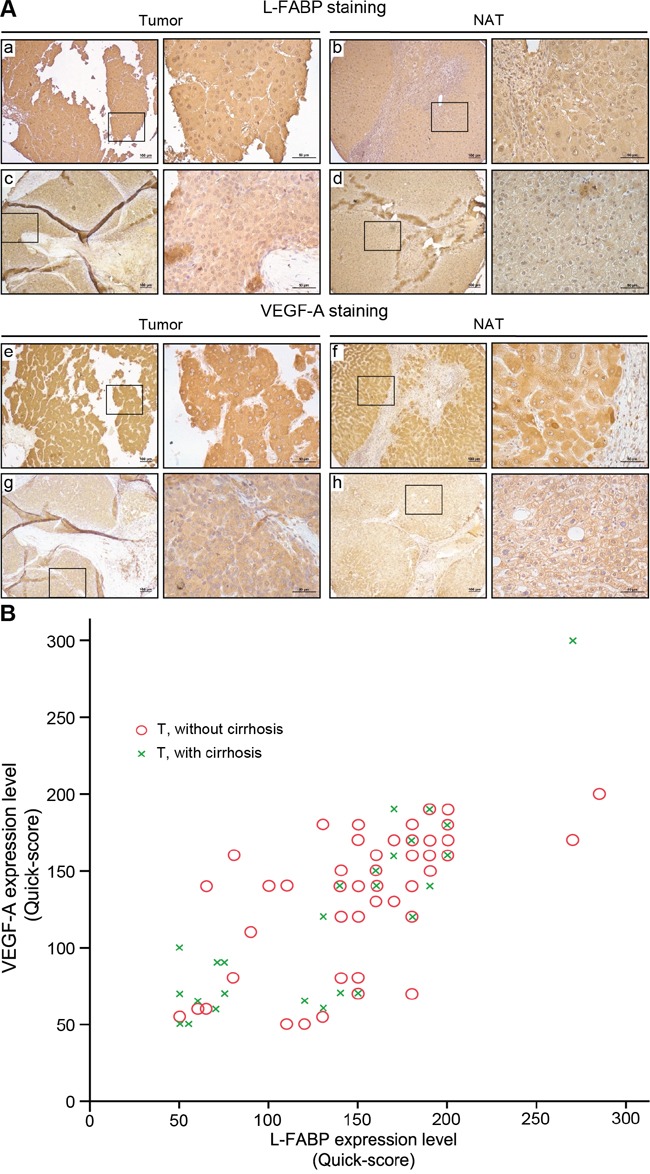
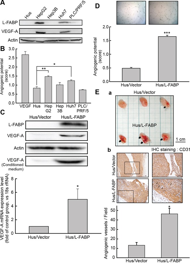
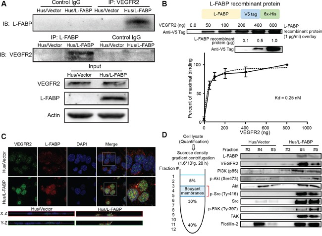
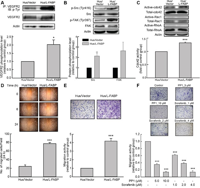
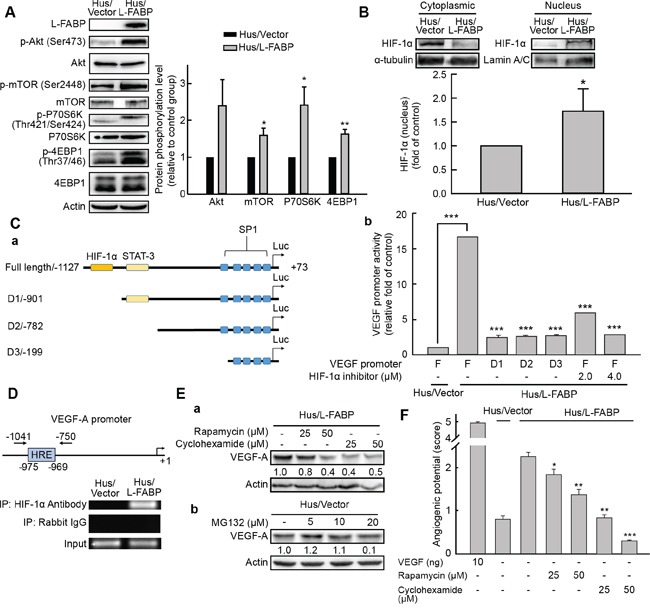
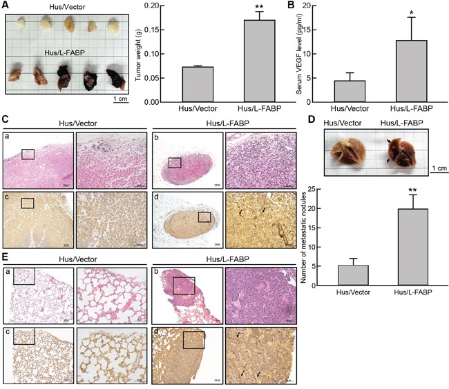
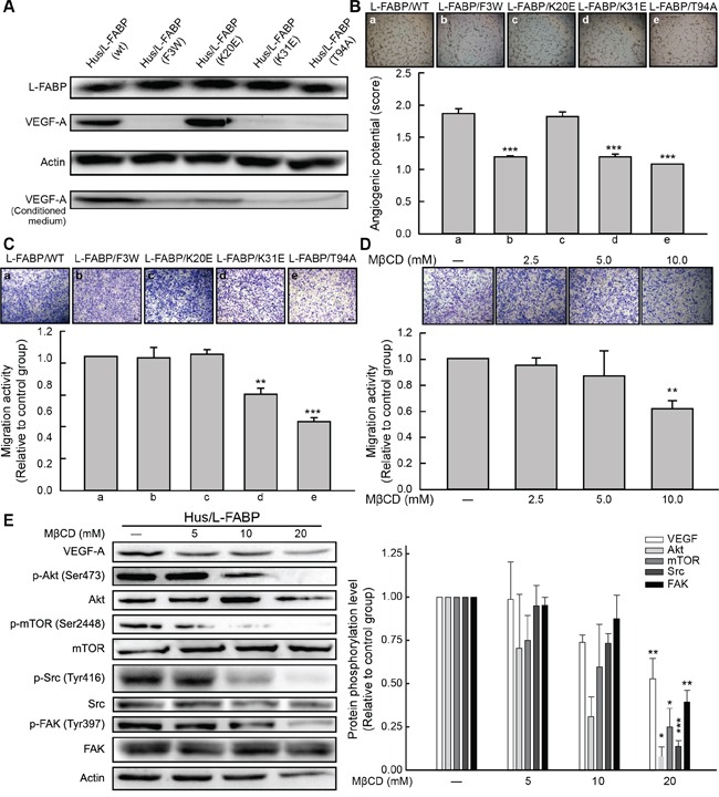
Liver fatty acid-binding protein (L-FABP) is abundant in hepatocytes and known to be involved in lipid metabolism. Overexpression of L-FABP has been reported in various cancers; however, its role in hepatocellular carcinoma (HCC) remains unclear. In this study, we investigated L-FABP and its association with vascular endothelial growth factors (VEGFs) in 90 HCC patients. We found that L-FABP was highly expressed in their HCC tissues, and that this expression was positively correlated with that of VEGF-A. Additionally, L-FABP significantly promoted tumor growth and metastasis in a xenograft mouse model. We also assessed the mechanisms of L-FABP activity in tumorigenesis; L-FABP was found to associate with VEGFR2 on membrane rafts and subsequently activate the Akt/mTOR/P70S6K/4EBP1 and Src/FAK/cdc42 pathways, which resulted in up-regulation of VEGF-A accompanied by an increase in both angiogenic potential and migration activity. Our results thus suggest that L-FABP could be a potential target for HCC chemotherapy.

Keywords: angiogenesis; hepatocellular carcinoma; liver fatty acid-binding protein; vascular endothelial growth factor.


Conflict of interest statement

CONFLICTS OF INTEREST

No conflict of interest exists in the submission of this manuscript, and the manuscript was approved by all authors for publication.

Figures

Figure 1
Figure 1. Expression of L-FABP and VEGF-A in tissues obtained from HCC patients
Protein expression was assessed in 90 HCC cases using immunohistochemical staining of paired normal (NAT) and tumor tissues. A. Staining of L-FABP and VEGF-A was observed in tumor tissues (L-FABP: a and c; VEGF-A: e and g) and their paired normal adjacent tissues (L-FABP: b and d; VEGF-A: f and h). Staining intensity: a and e, strong; b, c, f, and g, moderate; d and h, weak. B. Positive correlation between L-FABP and VEGF-A expression in 90 HCC tissues with and without cirrhosis (Pearson's correlation coefficient, r = 0.737; p < 0.01).
Figure 2
Figure 2. L-FABP promotes VEGF-A expression and angiogenic activity of liver cells
A. Western blot analysis for L-FABP expression in normal immortalized hepatocyte (Hus) and hepatocellular carcinoma (HepG2, Hep3B, Huh7 and PLC/PRF/5) cell lines. B. Angiogenic potential (score: see In vitro tube formation assay in Methods for details) of Hus, HepG2, Hep3B, Huh7, and PLC/PRF/5 cells. ***p < 0.001 versus Hus cells. C. Western blotting analysis of L-FABP and VEGF-A expression in Hus/L-FABP and Hus/Vector (vector-only control) cells. *p < 0.05 versus Hus/Vector control. D. In vitro angiogenic potential (score: see panel B) of Hus/L-FABP and Hus/Vector cells. Angiogenic vascular tube was imaged at 8 h. ***p < 0.001 versus Hus/Vector control. E. In vivo angiogenic activity of Hus/L-FABP and Hus/Vector cells assessed using a Matrigel plug assay. a: Matrigel plugs recovered from mice injected with Hus/Vector and Hus/L-FABP cells. Arrows indicate infiltration of blood vessels. b: Immunohistochemical (IHC) staining of CD31 (angiogenesis marker) in Matrigel plugs showed that Hus/L-FABP promoted angiogenesis, and the positively stained vessels are indicated by arrows. *p < 0.05 versus Hus/Vector control (n = 3).
Figure 3
Figure 3. L-FABP associates with VEGFR2 on membrane rafts
A. Left: Hus/L-FABP and Hus/Vector (vector-only control) cells were subjected to either immunoprecipitation (IP) with a VEGFR2 antibody followed by blotting with L-FABP or IP with an L-FABP antibody followed by blotting with VEGFR2. Right: Cell lysates (50 μg) were immunoblotted as an input control. B. L-FABP/V5-tagged recombinant protein was purified by Ni-NTA resin and subjected to SDS-PAGE to determine the purity as shown in the top photo. The overlay assay (far western blot analysis) was performed to estimate the affinity of the interaction between the VEGFR2 intracellular domain and L-FABP. PVDF membranes containing 0.5 to 8.0 μg of VEGFR2 recombinant protein (aa 789 to end) were incubated with L-FABP/V5-tagged recombinant protein (1 μg/ml) for 12 hours. The specific binding between L-FABP and VEGFR2 increased obviously between 0 to 2 μg, and maximal binding was observed at 8μg. The binding observed at the other concentrations was expressed as a percentage of the maximal binding within each experiment, and the Kd for binding between L-FABP and VEGFR2 was calculated as 0.25 nM. C. Three-color confocal images of cells that were fixed and stained with antibodies against L-FABP and VEGFR2. Signals: green, VEGFR2-Alexa 488; red, L-FABP-Alexa 568; and blue, DAPI. Magnification: 63×. The bottom photos show the X-Z and Y-Z optical sections, respectively, of Hus/Vector and Hus/L-FABP cells. Arrows indicate the co-localization of VEGFR2 and L-FABP in the apical membrane. D. Membrane localization of L-FABP, VEGFR2, PI3K (p85), phospho-Akt (Ser473), Akt, phosho-Src (Tyr416), Src, FAK, and phosho-FAK (Tyr397) in Hus/L-FABP or Hus/Vector cells. Membrane rafts were determined by using sucrose gradient-based ultracentrifugation and analyzed with western blotting (fraction #3–5).
Figure 4
Figure 4. L-FABP increases cell migration activity via VEGFR2/Src signaling and the FAK/cdc42 pathway
A. Phosphorylation of VEGFR2 in Hus/L-FABP and Hus/Vector (vector-only control) cells assessed by immunoprecipitation (IP) with a VEGFR2 antibody and blotting with a phospho-tyrosine antibody. *p < 0.05 versus Hus/Vector control. B. Phosphorylation of Src (Tyr416) and FAK (Tyr397) in Hus/L-FABP and Hus/Vector cells analyzed by western blotting. **p < 0.01 versus Hus/Vector control. C. Small GTPase binding in Hus/L-FABP or Hus/Vector cells. Active cdc42 and Rac1, but not RhoA, were detected by western blotting analysis. ***p < 0.001 for cdc42 activity versus Hus/Vector control. D. Wound-healing migration (2D migration activity) of Hus/L-FABP and Hus/Vector cells over 24 h. ***p < 0.001 versus Hus/Vector control. E. Migration activity of Hus/L-FABP and Hus/Vector cells seeded onto Boyden chambers and allowed to migrate toward 10% serum-containing medium for 16 h. ***p < 0.001 versus Hus/Vector control. F. Migration activity of Hus/L-FABP cells treated with PP1 (Src inhibitor: 5 or 10 μM) or Sorafenib (VEGFR2 inhibitor: 1, 2, or 4 μM) for 16 h. ***p < 0.001 versus DMSO-treated control group.
Figure 5
Figure 5. L-FABP-promoted VEGF-A expression is regulated by HIF-1α via the Akt/mTOR/P70S6K/4EBP1 pathway
A. Phosphorylation of Akt (Ser473), mTOR (Ser2448), P70S6K (Thr421/Ser424), and 4EBP1 (Thr37/46) in Hus/L-FABP and Hus/Vector (vector-only control) cells analyzed by western blotting. *p < 0.05 and **p < 0.01 versus Hus/Vector control. B. Nucleus and cytoplasmic localization of HIF-1α in Hus/L-FABP cells. Loading controls were α-tubulin and lamin A/C for cytoplasm and nucleus, respectively. HIF-1α levels increased ~1.7-fold in Hus/L-FABP relative to the control group: *p < 0.05. C. a: Diagram of the receptor constructs for the full-length VEGF-A promoter and deletion mutants (D1-D3). b: The luciferase activity of cell extracts was analyzed using a luciferase reporter assay (bar graph). ***p < 0.001 versus Hus/Vector control. D. Chromatin immunoprecipitation assay was performed to determine the amount of HIF-1α binding to the VEGF-A promoter; rabbit IgG served as a negative control, and the input served as a positive control. E. a: Western blot analysis of Hus/L-FABP cells treated with rapamycin (mTOR inhibitor: 25 or 50 μM) or cyclohexamide (translation inhibitor: 25 or 50 μM) for 12 h. b: Hus/Vector cells treated with MG132 (proteasome inhibitor: 5, 10, or 20 μM) for 24 h. F. In vitro angiogenic activity (score: see In vitro tube formation assay in Methods for details) of Hus/L-FABP and Hus/Vector cells treated with rapamycin or cyclohexamide (doses identical to those in D) for 12 h. ***p < 0.001 versus Hus/Vector control.
Figure 6
Figure 6. L-FABP promotes tumor growth and metastasis in vivo
Hus/L-FABP or Hus/Vector (vector-only control) cells (2 × 10⁶) was subcutaneously injected into the hind limbs of NOD/SCID mice, and the resulting in situ tumors were removed after 8 weeks for analysis. A. Representative photograph and average weight of tumors (n = 5 per group). **p < 0.01 versus Hus/Vector control. B. VEGF-A content in the serum of treated mice. *p < 0.001 versus Hus/Vector control. C. Stained tumor sections from Hus/L-FABP- and Hus/Vector-injected mice. H&E staining (a and b) and anti-CD31 antibody immunohistochemical staining (c and d). The positive staining indicated by the arrows shows strong angiogenic activity in the Hus/L-FABP-injected group. D. Metastatic activity of Hus/L-FABP and Hus/Vector cells (5 × 10⁶) in a lung metastasis model (NOD/SCID mice). After 10 weeks, the lungs were excised from mice (see photograph), and metastatic nodules (indicated by arrows) were counted (n = 5 per group). **p < 0.01 versus Hus/Vector control. E. Angiogenesis activity in metastatic nodules was assessed via H&E staining (a and b) or anti-CD31 immunohistochemical staining (c and d), and positive staining is indicated by arrows.
Figure 7
Figure 7. Cholesterol binding properties are essential for L-FABP-induced cell migration and angiogenesis
A. Western blotting analysis of L-FABP and VEGF-A expression (both intracellular and extracellular levels) in various mutants of L-FABP-overexpressed stable cells generated by site-directed mutagenesis B. In vitro angiogenic activity (score: see In vitro tube formation assay in Methods for details) of mutants. Images represent amino acid substitutions: (a) L-FABP (wild type), (b) L-FABP (F3 to W), (c) L-FABP (K20 to E), (d) L-FABP (K31 to E), and (e) L-FABP (T94 to A). ***p < 0.001 versus wild-type. C. Migration activity of the mutants. Images (a–e) represent the amino acid substitutions described in (B). **p < 0.01, ***p < 0.001 versus wild-type. D. Migration activity of Hus/L-FABP cells treated with MβCD (cholesterol depletion agent: 5, 10, or 20 mM) for 12 h. **p < 0.01 versus water-treated control group. E. Western blot analysis of Hus/L-FABP cells treated with MβCD (5, 10, or 20 mM) for 6 h. *p < 0.05, **p < 0.01, ***p < 0.001 versus water-treated control group.

Similar articles

Cited by

References

    1. Kaseb AO, Hanbali A, Cotant M, Hassan MM, Wollner I, Philip PA. Vascular endothelial growth factor in the management of hepatocellular carcinoma: a review of literature. Cancer. 2009;115:4895–4906. - PubMed
    1. Moeini A, Cornellà H, Villanueva A. Emerging signaling pathways in hepatocellular carcinoma. Liver Cancer. 2012;1:83–93. - PMC - PubMed
    1. Romanque P, Piguet AC, Dufour JF. Targeting vessels to treat hepatocellular carcinoma. Clin Sci (Lond) 2008;114:467–477. - PubMed
    1. Zhu AX, Duda DG, Sahani DV, Jain RK. HCC and angiogenesis: possible targets and future directions. Nat Rev Clin Oncol. 2011;8:292–301. - PMC - PubMed
    1. Storch J, McDermott L. Structural and functional analysis of fatty acid-binding proteins. J Lipid Res. 2009;50:S126–131. - PMC - PubMed

Publication types

MeSH terms

Substances