. 2017 Mar;27(2):146-159.
doi: 10.1111/bpa.12367. Epub 2016 Apr 19.

Molecular Diagnostics of Gliomas Using Next Generation Sequencing of a Glioma-Tailored Gene Panel

Affiliations

Molecular Diagnostics of Gliomas Using Next Generation Sequencing of a Glioma-Tailored Gene Panel

Angela Zacher et al. Brain Pathol. 2017 Mar.

Abstract

Current classification of gliomas is based on histological criteria according to the World Health Organization (WHO) classification of tumors of the central nervous system. Over the past years, characteristic genetic profiles have been identified in various glioma types. These can refine tumor diagnostics and provide important prognostic and predictive information. We report on the establishment and validation of gene panel next generation sequencing (NGS) for the molecular diagnostics of gliomas. We designed a glioma-tailored gene panel covering 660 amplicons derived from 20 genes frequently aberrant in different glioma types. Sensitivity and specificity of glioma gene panel NGS for detection of DNA sequence variants and copy number changes were validated by single gene analyses. NGS-based mutation detection was optimized for application on formalin-fixed paraffin-embedded tissue specimens including small stereotactic biopsy samples. NGS data obtained in a retrospective analysis of 121 gliomas allowed for their molecular classification into distinct biological groups, including (i) isocitrate dehydrogenase gene (IDH) 1 or 2 mutant astrocytic gliomas with frequent α-thalassemia/mental retardation syndrome X-linked (ATRX) and tumor protein p53 (TP53) gene mutations, (ii) IDH mutant oligodendroglial tumors with 1p/19q codeletion, telomerase reverse transcriptase (TERT) promoter mutation and frequent Drosophila homolog of capicua (CIC) gene mutation, as well as (iii) IDH wildtype glioblastomas with frequent TERT promoter mutation, phosphatase and tensin homolog (PTEN) mutation and/or epidermal growth factor receptor (EGFR) amplification. Oligoastrocytic gliomas were genetically assigned to either of these groups. Our findings implicate gene panel NGS as a promising diagnostic technique that may facilitate integrated histological and molecular glioma classification.

Keywords: glioma; molecular diagnostics; mutation; next generation sequencing.


Figures

Figure 1
Figure 1
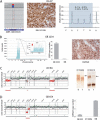
Examples of sequence and copy number changes detected by gene panel NGS and validated by independent methods. A. Detection of IDH1‐R132H mutation in an oligoastrocytoma (OA 97). Shown are selected IDH1 sequencing reads from a total number of 2911 reads, with 44% of all reads corresponding to C>T transition translating into IDH1‐R132H (left). Presence of the IDH1 mutation in OA 97 was validated by immunohistochemistry (middle) and DNA pyrosequencing (right), the latter technique demonstrating a mutant allele frequency of 47%. B. Detection of EGFR amplification and the EGFRvIII deletion rearrangement by gene panel NGS in a glioblastoma (GB 2234). EGFR amplification is reflected by a strong increase in the number of total reads obtained for the EGFR sequences relative to all other sequences (left). The EGFRvIII deletion rearrangement is indicated by reduced read counts obtained for EGFR exons 2‐7 (red arrow) relative to the read counts for the highly amplified EGFR exons 1 and 8–28 (left, insert). EGFR amplification in GB 2234 was confirmed by quantitative real‐time PCR (middle; NB, normal brain). Presence of EGFRvIII was validated by immunohistochemical staining with an EGFRvIII‐specific antibody (right). C. Detection of 1p/19q codeletion in an anaplastic oligodendroglioma (AO 84) by demonstrating reduced coverage ratios for gene sequences on 1p and 19q (left, red data points). Validation of the 1p/19q codeletion in AO 84 was performed by microsatellite analysis (right) demonstrating loss of heterozygosity at microsatellite markers D1S2696 (1p) and D19S219 (19q) in tumor (T) DNA (arrows) relative to the corresponding blood DNA (B). D. Detection of a homozygous CDKN2A deletion in a glioblastoma (GB 654) by gene panel NGS (left). Note that gene sequences on 9p21 (CDKN2A) and 10q23 (PTEN) demonstrate reduced coverage ratios, with the coverage ratio for CDKN2A amplicons (homozygous deletion) being even lower than for PTEN amplicons (hemizygous deletion). Homozygous CDKN2A deletions were validated by quantitative PCR using a CDKN2A TaqMan® copy number assays (right). Normal brain (NB) DNA was used as constitutive control with two gene copies, while U118MG DNA served as positive control for homozygous deletion.
Figure 2
Figure 2
Results of unsupervised hierarchical cluster analysis of the 20‐gene panel NGS data obtained in 121 gliomas. Blue and gray boxes indicate presence or absence of a given aberration. Note that tumors form major clusters primarily characterized by frequent mutations in IDH1 or H3F3A, TP53 and/or ATRX (green bar), frequent mutations in IDH1 or IDH2, the TERT promoter and CIC, as well as 1p/19q codeletion (red bar), or frequent mutations in the TERT promoter, PTEN and/or NF1, as well as EGFR amplification (blue bar). The remaining tumors (gray bar) included a subgroup of BRAF‐V600E mutant gliomas as well as another subgroup of tumors without detectable aberrations or only single gene mutations. Histological classification of the individual tumors according to the WHO classification 2007 is indicated (A II, astrocytoma WHO grade II; AA III, anaplastic astrocytoma WHO grade III; O II, oligodendroglioma WHO grade II; AO III, anaplastic oligodendroglioma WHO grade III; OA II, oligoastrocytoma WHO grade II; AOA III, anaplastic oligoastrocytoma WHO grade III; GB IV, glioblastoma WHO grade IV; PA I, pilocytic astrocytoma WHO grade I; PXA, pleomorphic xanthoastrocytoma); *includes TERT mutations detected by gene panel NGS and/or conventional sequencing; **refers to CDKN2A homozygous deletion:***includes EGFRvIII mutations detected by gene panel NGS and/or immunohistochemistry/RT‐PCR.
Figure 3
Figure 3
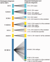
Schematic illustration of the diagnostic changes in 111 diffusely infiltrative gliomas originally classified histologically according to the WHO classification of 2007 (left side) and then reclassified by integrating histology and the genetic markers IDH mutation, 1p/19q codeletion and H3F3A‐K27M mutation in line with the Haarlem consensus recommendations 30 for the upcoming revised WHO classification. *The three H3F3A‐K27 mutant tumors were all midline (thalamic) lesions.

Similar articles

Cited by

References

    1. Adzhubei IA, Schmidt S, Peshkin L, Ramensky VE, Gerasimova A, Bork P et al (2010) A method and server for predicting damaging missense mutations. Nat Methods 7:248–249. - PMC - PubMed
    1. Appin CL, Brat DJ (2015) Molecular pathways in gliomagenesis and their relevance to neuropathologic diagnosis. Adv Anat Pathol 22:50–58. - PubMed
    1. Bechet D, Gielen, Gerrit G H, Korshunov A, Pfister SM, Rousso C et al (2014) Specific detection of methionine 27 mutation in histone 3 variants (H3K27M) in fixed tissue from high‐grade astrocytomas. Acta Neuropathol 128:733–741. - PMC - PubMed
    1. Brat DJ, Verhaak, Roel G W, Aldape KD, Yung WKA, Salama SR et al (2015) Comprehensive, integrative genomic analysis of diffuse lower‐grade gliomas. N Engl J Med 372:2481–2498. - PMC - PubMed
    1. Brennan CW, Verhaak RGW, McKenna A, Campos B, Noushmehr H, Salama SR, Zheng S et al (2013) The somatic genomic landscape of glioblastoma. Cell 155:462–477. - PMC - PubMed

Publication types

MeSH terms

Supplementary concepts