Ab initio structure determination of nanocrystals of organic pharmaceutical compounds by electron diffraction at room temperature using a Timepix quantum area direct electron detector
- PMID: 26919375
- PMCID: PMC4770873
- DOI: 10.1107/S2053273315022500
Ab initio structure determination of nanocrystals of organic pharmaceutical compounds by electron diffraction at room temperature using a Timepix quantum area direct electron detector
Erratum in
-
Ab initio structure determination of nanocrystals of organic pharmaceutical compounds by electron diffraction at room temperature using a Timepix quantum area direct electron detector. Corrigendum.Acta Crystallogr A Found Adv. 2018 Nov 1;74(Pt 6):709. doi: 10.1107/S2053273318014079. Epub 2018 Oct 30. Acta Crystallogr A Found Adv. 2018. PMID: 30378582 Free PMC article.
Abstract
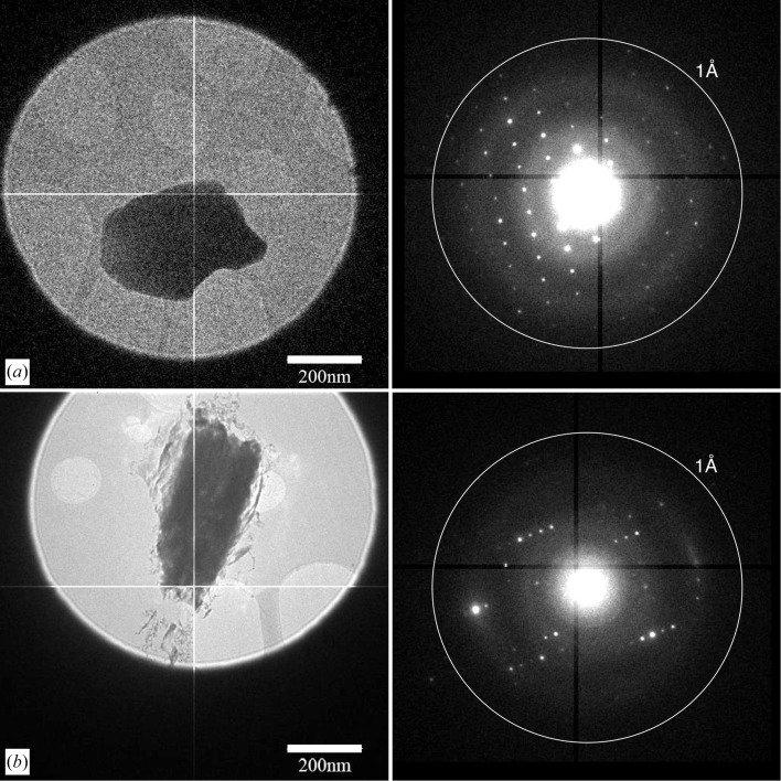
Until recently, structure determination by transmission electron microscopy of beam-sensitive three-dimensional nanocrystals required electron diffraction tomography data collection at liquid-nitrogen temperature, in order to reduce radiation damage. Here it is shown that the novel Timepix detector combines a high dynamic range with a very high signal-to-noise ratio and single-electron sensitivity, enabling ab initio phasing of beam-sensitive organic compounds. Low-dose electron diffraction data (∼ 0.013 e(-) Å(-2) s(-1)) were collected at room temperature with the rotation method. It was ascertained that the data were of sufficient quality for structure solution using direct methods using software developed for X-ray crystallography (XDS, SHELX) and for electron crystallography (ADT3D/PETS, SIR2014).
Keywords: Timepix quantum area detector; carbamazepine; electron diffraction structure determination; electron nanocrystallography; nicotinic acid.
Figures
References
-
- Abrahams, J. P. (1993). Jnt CCP4/ESF-EACBM Newsl. Protein Crystallogr. 28, 3–4.
-
- Arndt, W. & Wonacott, A. J. (1977). Editors. The Rotation Method in Crystallography. Amsterdam: North-Holland.
-
- Brönnimann, E., Baur, R., Eikenberry, E. F., Fischer, P., Florin, S., Horisberger, R., Lindner, M., Schmitt, B. & Schulze, C. (2002). Nucl. Instrum. Methods Phys. Res. Sect. A, 477, 531–535.
-
- Bruker (2004). SAINT-Plus and XPREP. Bruker Axs Inc., Madison, Wisconsin, USA.
-
- Burla, M. C., Caliandro, R., Carrozzini, B., Cascarano, G. L., Cuocci, C., Giacovazzo, C., Mallamo, M., Mazzone, A. & Polidori, G. (2015). J. Appl. Cryst. 48, 306–309.
Publication types
MeSH terms
Substances
LinkOut - more resources
Full Text Sources
Other Literature Sources