

Cut to the chase: a review of CD26/dipeptidyl peptidase-4's (DPP4) entanglement in the immune system
- PMID: 26919392
- PMCID: PMC4908298
- DOI: 10.1111/cei.12781
Cut to the chase: a review of CD26/dipeptidyl peptidase-4's (DPP4) entanglement in the immune system
Abstract
CD26/DPP4 (dipeptidyl peptidase 4/DP4/DPPIV) is a surface T cell activation antigen and has been shown to have DPP4 enzymatic activity, cleaving-off amino-terminal dipeptides with either L-proline or L-alanine at the penultimate position. It plays a major role in glucose metabolism by N-terminal truncation and inactivation of the incretins glucagon-like peptide-1 (GLP) and gastric inhibitory protein (GIP). In 2006, DPP4 inhibitors have been introduced to clinics and have been demonstrated to efficiently enhance the endogenous insulin secretion via prolongation of the half-life of GLP-1 and GIP in patients. However, a large number of studies demonstrate clearly that CD26/DPP4 also plays an integral role in the immune system, particularly in T cell activation. Therefore, inhibition of DPP4 might represent a double-edged sword. Apart from the metabolic benefit, the associated immunological effects of long term DPP4 inhibition on regulatory processes such as T cell homeostasis, maturation and activation are not understood fully at this stage. The current data point to an important role for CD26/DPP4 in maintaining lymphocyte composition and function, T cell activation and co-stimulation, memory T cell generation and thymic emigration patterns during immune-senescence. In rodents, critical immune changes occur at baseline levels as well as after in-vitro and in-vivo challenge. In patients receiving DPP4 inhibitors, evidence of immunological side effects also became apparent. The scope of this review is to recapitulate the role of CD26/DPP4 in the immune system regarding its pharmacological inhibition and T cell-dependent immune regulation.
Keywords: B cell; T cells; autoimmunity; cell activation; chemokines.
© 2016 British Society for Immunology.
Figures
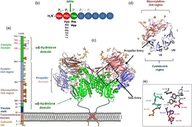
 , N‐glycosylation;
  , potential unoccupied N‐glycosylation;
  , cysteine residues involved in S‐bridges; red numbers and letters indicate the catalytic triad. (b) Substrate specificity of DPP4. Xaa and Yaa indicate any amino acid. Decreasing font of amino acid at P1 position represents declining rate of hydrolysis. Amino acids crossed out must not occupy P1′. Arrow indicates site of cleavage. (c) quaternary structure of homodimeric human recombinant DPP4 as determined by Weihofen et al., 2004, showing the α/β‐hydrolase domain (aa 39–51 and 506–766) in green and propeller domain (aa 55–497) with the glycosylation‐rich subdomain (red) and the cystein‐rich subdomain (blue). (d) Propeller domain viewed from the top, illustrating the eight propeller blades designated with roman numbers and two subdomains. (e) Active site zoomed in, depicting the residues involved in catalysis, catalytic triad Ser630, Asp708, His740 are shown in red, Tyr547 responsible for oxyanion hole in brown, Tyr662 and Tyr666 forming the hydrophobic pocket in grey, Arg125 and Asn710, contributing to an electrostatic sink in orange and blue, respectively, and Glu205 and Glu206 ensuring N‐terminal anchoring in pale green. S–S bridges are illustrated in yellow and carbohydrates in orange. Structures were drawn with PyMOLTM 2008 DeLano Scientific LLC, using Protein Data Base: 1W1I 7.
References
-
- Hopsu‐Havu VK, Glenner GG. A new dipeptide naphthylamidase hydrolyzing glycyl‐prolyl‐beta‐naphthylamide. Histochemie 1966; 7:197–201. - PubMed
-
- De Meester I, Korom S, Van Damme J, Scharpé S. CD26, let it cut or cut it down. Immunol Today 1999; 20:367–75. - PubMed
-
- Lambeir A‐M, Durinx C, Scharpé S, De Meester I. Dipeptidyl‐peptidase IV from bench to bedside: an update on structural properties, functions, and clinical aspects of the enzyme DPP IV. Crit Rev Clin Lab Sci 2003; 40:209–94. - PubMed
-
- Gorrell MD. Dipeptidyl peptidase IV and related enzymes in cell biology and liver disorders. Clin Sci (Lond) 2005; 108:277–92. - PubMed
Publication types
MeSH terms
Substances
LinkOut - more resources
Full Text Sources
Other Literature Sources
Medical
Miscellaneous
