. 2016 Feb 25;164(5):896-910.
doi: 10.1016/j.cell.2015.12.057.

NF-κB Restricts Inflammasome Activation via Elimination of Damaged Mitochondria

Affiliations

NF-κB Restricts Inflammasome Activation via Elimination of Damaged Mitochondria

Zhenyu Zhong et al. Cell. .

Abstract

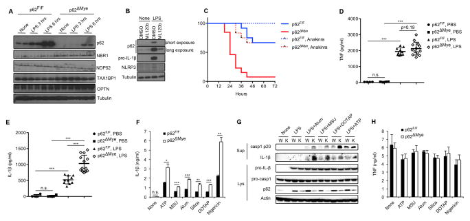
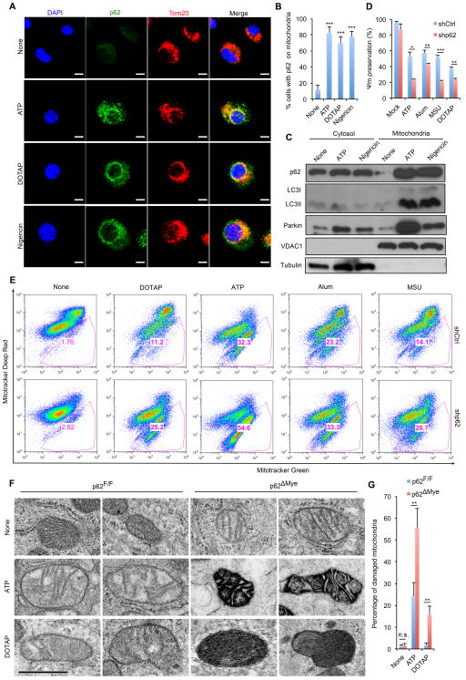
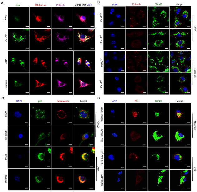
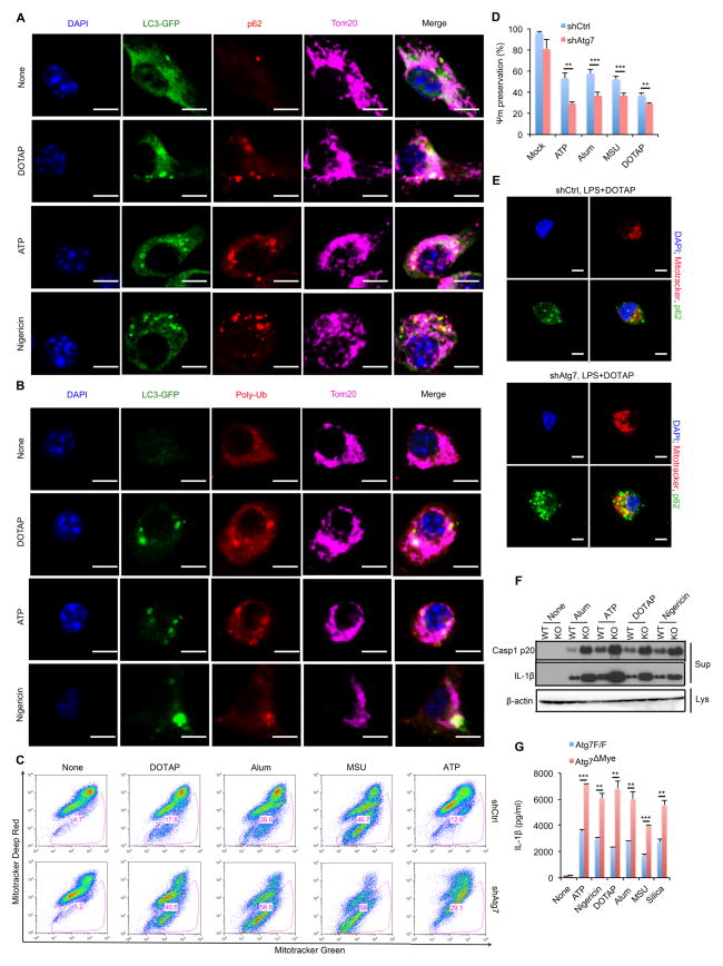
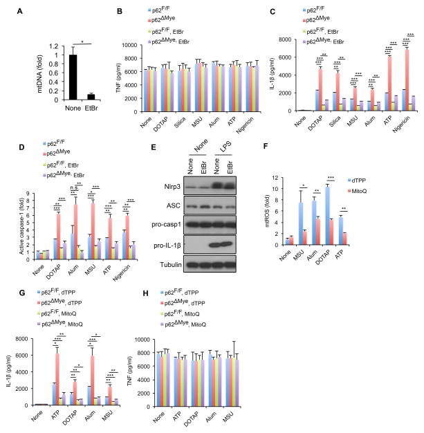
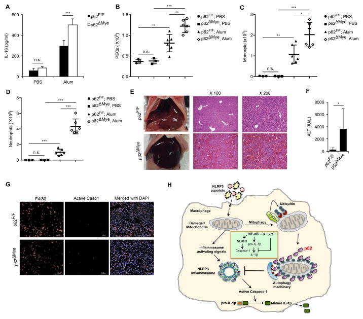
Nuclear factor κB (NF-κB), a key activator of inflammation, primes the NLRP3-inflammasome for activation by inducing pro-IL-1β and NLRP3 expression. NF-κB, however, also prevents excessive inflammation and restrains NLRP3-inflammasome activation through a poorly defined mechanism. We now show that NF-κB exerts its anti-inflammatory activity by inducing delayed accumulation of the autophagy receptor p62/SQSTM1. External NLRP3-activating stimuli trigger a form of mitochondrial (mt) damage that is caspase-1- and NLRP3-independent and causes release of direct NLRP3-inflammasome activators, including mtDNA and mtROS. Damaged mitochondria undergo Parkin-dependent ubiquitin conjugation and are specifically recognized by p62, which induces their mitophagic clearance. Macrophage-specific p62 ablation causes pronounced accumulation of damaged mitochondria and excessive IL-1β-dependent inflammation, enhancing macrophage death. Therefore, the "NF-κB-p62-mitophagy" pathway is a macrophage-intrinsic regulatory loop through which NF-κB restrains its own inflammation-promoting activity and orchestrates a self-limiting host response that maintains homeostasis and favors tissue repair.


Figures

Figure 1
Figure 1. p62, induced on NF-κB activation, suppresses NLRP3-inflammasome dependent IL-1β production
(A) Immunoblot (IB) analysis of autophagy receptors in wild-type (p62F/F) and p62ΔMye BMDM stimulated with LPS (200 ng/ml). (B) Wild-type BMDM were incubated with or without LPS in the absence or presence of IKKβ inhibitor ML120b and expression of p62, pro-IL-1β, and NLRP3 was IB analyzed. (C) Survival of p62F/F or p62ΔMye mice injected with intraperitoneal (i.p.) LPS (30 mg/kg) without or with anakinra (50 mg/kg), n=11–13. (D, E) 12-weeks old p62F/F or p62ΔMye mice were i.p. injected with LPS and their sera collected 3 hrs later and analyzed by ELISA for TNF (D) and IL-1β (E). (F, H) Release of IL-1β (F) or TNF (H) from LPS-primed p62F/F or p62ΔMye BMDM that were stimulated with the indicated NLRP3 agonists. Data are averages ± s.d. (n=3). (G) IB analysis of caspase-1 and pro-IL-1β processing in LPS-primed WT (W) or p62-deficient (K) iBMDM stimulated as indicated. Data are representative of three independent experiments. *, p<0.05; **, p<0.01; ***, p<0.001.
Figure 2
Figure 2. NLRP3 agonists promote mitochondrial damage and p62 recruitment
(A) Intracellular distribution of p62 and mitochondria (Tom20) in LPS-primed BMDM stimulated with NLRP3 agonists examined by confocal microscopy (Scale bars, 5 μm) and (B) quantitated by counting cells with p62 aggregation on mitochondria. Averages ± s.d. represent the ratio of cells with mitochondrial p62 aggregates to total cells in each field. (n=6). (C) Subcellular distribution of p62, LC3II, and Parkin before and after NLRP3-inflammasome activation. Data are representative of three independent experiments. (D) NLRP3 agonist-induced changes in mitochondrial membrane potential (Ψm) in LPS-primed WT (shCtrl) or p62-deficient (shp62) iBMDM were measured by TMRM fluorescence. Data are averages ± s.d.(n=3). (E) Flow cytometric analysis of mitochondrial status in macrophages challenged with NLRP3 agonists. Gates represent cells with damaged mitochondria. (F, G) Electron micrographs of mitochondria in LPS-primed p62F/F or p62ΔMye BMDM after incubation with NLRP3 agonists. Shown are representative examples of normal, partially damaged and heavily damaged mitochondria. Scale bars: 500 nm. (G) quantification of damaged mitochondria in (F). Results are averages ± s.e.m. (n=8). *, p<0.05; **, p<0.01; ***, p<0.001.
Figure 3
Figure 3. Mitochondrial p62 recruitment is Parkin dependent
(A) Intracellular distribution of p62, polyubiquitin (poly-Ub) and mitochondria (Mitotracker) in LPS-primed WT BMDM stimulated with NLRP3 agonists determined by confocal microscopy. Scale bars: 5 μm. (B) Mitochondrial poly-Ub decoration examined by confocal microscopy in LPS-primed Park2+/+ or Park2−/− BMDM stimulated with NLRP3 agonists. Scale bars: 5 μm. (C) Mitochondrial recruitment of p62 in LPS-primed WT (shCtrl) or Parkin-deficient (shPark2) iBMDM stimulated with NLRP3 agonists. Scale bars: 5 μm. (D) Subcellular distribution of human p62 in LPS-primed, NLRP3 agonist stimulated p62-deficient BMDMs transduced with human p62-full length or p62(ΔUBA) constructs. Scale bars: 5 μm.
Figure 4
Figure 4. Parkin limits accumulation of damaged mitochondria after inflammasome activation
(A) Flow cytometry of mitochondrial status in WT (shCtrl) or Parkin-deficient (shPark2) iBMDM stimulated with NLRP3 agonists. Gates represent cells with damaged mitochondria. (B) NLRP3 agonist-induced changes in mitochondrial membrane potential (Ψm) in LPS-primed WT (shCtrl) or p62-deficient (shp62) iBMDM measured by TMRM fluorescence. Data are averages ± s.d. (n=3). (C) IL-1β release by LPS-primed Park2+/+ or Park2−/− BMDM stimulated with NLRP3 agonists. Data are averages ± s.d. (n=3). (D) Caspase-1 activity in LPS-primed WT (shCtrl) or Parkin-deficient (shPark2) iBMDM stimulated with NLRP3 agonists. Results are averages ± s.d. (n=3). (E) IB analysis of cleaved caspase-1 and IL-1β secretion to cell culture supernatants (Sup) or tubulin in cell lysates (Lys) in LPS-primed Park2+/+ or Park2−/− BMDM stimulated with NLRP3 agonists. Data are representative of 3 independent experiments. *, p<0.05; **, p<0.01; ***, p<0.001.
Figure 5
Figure 5. Autophagy mediates clearance of p62-bound mitochondria
(A, B) Intracellular distribution of LC3, p62, poly-Ub and mitochondria in LPS-primed LC3-GFP BMDM stimulated with NLRP3 agonists examined by confocal microscopy. (C) Flow cytometric analysis of mitochondrial status in LPS-primed WT (shCtrl) or Atg7-deficient (shAtg7) iBMDM after NLRP3 agonist stimulation. Gates – cells with damaged mitochondria. (D) NLRP3 agonist-induced changes in mitochondrial membrane potential (Ψm) in LPS-primed WT (shCtrl) or Atg7-deficient (shAtg7) iBMDM. Data are averages ± s.d. (n=3). (E) Intracellular distribution of p62 and mitochondria in LPS-primed shCtrl or shAtg7 iBMDM stimulated with NLRP3 agonists. (F) IB analysis of caspase-1 p20 and mature IL-1β in culture supernatants (Sup) and β-actin in cell lysates (Lys) of LPS-primed Atg7F/F (WT) and Atg7ΔMye (KO) BMDM stimulated as indicated. Data are representative of three independent experiments. (G) IL-1β secretion by above cells measured by ELISA. Results are averages ± s.d. (n=3). *, p<0.05; **, p<0.01; ***, p<0.001.
Figure 6
Figure 6. Elimination of mitochondrial signals prevents excessive IL-1β production by p62-deficient macrophages
(A) Relative concentrations of mtDNA in p62F/F and p62ΔMye BMDM before and after 4 days of ethidium bromide (EtBr) treatment. Results are averages ± s.d. (n=3). (B–D) TNF (B) IL-1β (C) release and caspase-1 activity (D) in p62F/F and p62ΔMye BMDM pre-treated with EtBr or not that were co-stimulated with LPS and different NLRP3 agonists. Results are averages ± s.d. (n=3). (E) IB analysis of pro-IL-1β, NLRP3, ASC, and pro-caspase-1 in lysates of EtBr-pretreated WT BMDM before and after 4 hrs of LPS stimulation. Data are representative of three independent experiments. (F) Relative mtROS amounts determined by MitoSOX staining of LPS-primed WT BMDM stimulated with NLRP3 agonists in the presence of dTPP or Mito-Q. (G,H) Release of IL-1β (F) and TNF (G) from LPS-primed p62F/F and p62ΔMye BMDM; treated with Mito-Q or vehicle (dTPP) before addition of NLRP3 agonists. Results are averages ± s.d. (n=3).
Figure 7
Figure 7. p62 inhibits inflammasome-dependent sterile inflammation
(A) Peritoneal IL-1β in p62F/F or p62ΔMye mice 4 hrs after i.p. injection of alum or PBS. n=3–6. (B–D) Alum-induced peritoneal infiltration of PEC (B), monocytes (CD11b+Ly6C+Ly6G) (C) and neutrophils (CD11b+Ly6G+F4/80) (D) in p62F/F and p62ΔMye mice 16 hrs after alum or PBS injection. (E) Representative liver appearance and histology of p62F/F or p62ΔMye mice i.p. injected with LPS plus D-galactosamine. (F) Serum ALT in above mice. (G) Fluorescent staining of F4/80, active caspase-1, and DAPI in livers of p62F/F and p62ΔMye mice after LPS+ D-gal challenge. *, p<0.05; **, p<0.01; ***, p<0.001. (H) Schematic representation of key findings. NF-κB induces expression of p62, which negatively regulates caspase-1 activation via mitophagic elimination of mitochondria that release NLRP3-inflammasome activating signals.

Comment in

References

    1. Barnes PJ, Karin M. Nuclear factor-kappaB: a pivotal transcription factor in chronic inflammatory diseases. N Engl J Med. 1997;336:1066–1071. - PubMed
    1. Baroja-Mazo A, Martin-Sanchez F, Gomez AI, Martinez CM, Amores-Iniesta J, Compan V, Barbera-Cremades M, Yague J, Ruiz-Ortiz E, Anton J, et al. The NLRP3 inflammasome is released as a particulate danger signal that amplifies the inflammatory response. Nat Immunol. 2014;15:738–748. - PubMed
    1. Brenner C, Galluzzi L, Kepp O, Kroemer G. Decoding cell death signals in liver inflammation. J Hepatol. 2013;59:583–594. - PubMed
    1. Brydges SD, Broderick L, McGeough MD, Pena CA, Mueller JL, Hoffman HM. Divergence of IL-1, IL-18, and cell death in NLRP3 inflammasomopathies. J Clin Invest. 2013;123:4695–4705. - PMC - PubMed
    1. Chernyak BV, Izyumov DS, Lyamzaev KG, Pashkovskaya AA, Pletjushkina OY, Antonenko YN, Sakharov DV, Wirtz KW, Skulachev VP. Production of reactive oxygen species in mitochondria of HeLa cells under oxidative stress. Biochim Biophys Acta. 2006;1757:525–534. - PubMed

Publication types

MeSH terms