Review
. 2017:404:1-44.
doi: 10.1007/82_2015_5021.

Envelope Structures of Gram-Positive Bacteria

Affiliations
Review

Envelope Structures of Gram-Positive Bacteria

Mithila Rajagopal et al. Curr Top Microbiol Immunol. 2017.

Abstract

Gram-positive organisms, including the pathogens Staphylococcus aureus, Streptococcus pneumoniae, and Enterococcus faecalis, have dynamic cell envelopes that mediate interactions with the environment and serve as the first line of defense against toxic molecules. Major components of the cell envelope include peptidoglycan (PG), which is a well-established target for antibiotics, teichoic acids (TAs), capsular polysaccharides (CPS), surface proteins, and phospholipids. These components can undergo modification to promote pathogenesis, decrease susceptibility to antibiotics and host immune defenses, and enhance survival in hostile environments. This chapter will cover the structure, biosynthesis, and important functions of major cell envelope components in gram-positive bacteria. Possible targets for new antimicrobials will be noted.


Figures
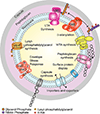
Fig 1
Fig 1
The Gram-positive cell envelope. The complex Gram-positive cell envelope is the first line of defense for the organism. Here, the S. aureus envelope is shown as an example. Major pathways involved in the synthesis of the cell envelope include capsule, PG and TA synthesis. TAs can be modified by d-alanlyation. d-alanylation and lysylphosphatidylglycerol synthesis are known factors for antibiotic resistance. Envelope stress response regulators modulate the organism's response to toxic molecules or conditions that perturb the cell envelope. Importers and exporters, ubiquitously present among bacteria, serve the necessary role of channeling in nutrients and pumping out toxic molecules. Finally, surface protein display systems function to tether proteins to the cell membrane or cell wall, which perform important roles in adhesion and interaction with the environment.
Fig 2
Fig 2
PG structure, and common variations. PG consists of chains of alternating GlcNAc and MurNAc residues. The MurNAc residues are functionalized with pentapeptide units which are cross-linked via the substituents on l-Lys to generate the mature PG. The linear glycan chain is highly conserved across both Gram-positives and Gram-negatives. The stem pentapeptide is well conserved across Gram-positives, aside from B. subtilis which contains meso-diaminopimmelic acid instead of l-Lysine at position 3 of the stem pentapeptide. There is considerable variation in the substituents on the l-Lys across Gram-positive species as indicated. PG can be modified by O-acetylation of MurNAc or N-deacetylation of GlcNAc moieties in response to challenge from antimicrobials such as lysozyme.
Fig 3
Fig 3
Synthesis of PG and antibiotics that target PG synthesis. The enzymatic steps for PG synthesis are well conserved across species. Here, the biosynthesis of S. aureus PG is shown as an example. The synthesis begins with the assembly of the GlcNAc-MurNAc-pentapeptide and its attachment to carrier lipid Und-P in the cell membrane. After this point, the l-Lysine at position 3 is substituted with additional amino acids and then flipped to the outside of the cell where it is cross-linked by PBPs. The same lipid carrier is also utilized for WTA (shown here) and capsule synthesis. The synthesis of PG is crucial to the cell and over time, several antibiotics have been discovered that target various steps in PG biosynthesis.
Fig 4
Fig 4
WTA structure, and common variations. WTAs are anionic polymers with a sugar-phosphate backbone attached to the C6 position of MurNAc in PG. The structure of WTAs is highly variable across Gram-positive species. WTA polymer structures for specific strains are indicated here with the following abbreviations: Glycerol-phosphate (GroP), Ribitol Phosphate (RboP), N-acetylmannosamine (ManNAc), Galactose (Gal), Glucose (Gluc), N-acetylgalactosamine (GalNAc), N-acetylglucosamine (GlcNAc), phosphorylcholine (choline-P).
Fig 5
Fig 5
TA biosynthesis and modification pathways. LTA and WTA biosynthetic pathways in S. aureus are shown here. Although both are anionic sugar-phosphate backbones, they are assembled differently by separate biosynthetic pathways in S. aureus. TAs are further modified with d-alanine residues by the dlt pathway and with α- or β- GlcNAC residues installed by glycosyltransferases TarM and TarS, respectively. TAs perform several functions for the cell including playing roles in biofilm formation, adhesion, phage attachment, virulence and antibiotic resistance, most notably resistance to β-lactams. d-alanylation has been shown to play an important role in these functions as well. Specifically, the absence of d-alanine modifications sensitizes to cationic antimicrobial peptides, including host defensins. The only known roles for α- and β- GlcNAC modifications are in phage attachment, and for β-GlcNAcs, in β-lactam resistance. Due to its roles in adhesion, virulence and antibiotic resistance, attempts are being made to target TA biosynthesis and modification pathways. The known compounds targeting these pathways are shown here.

References

    1. Abachin E, Poyart C, Pellegrini E, Milohanic E, Fiedler F, Berche P, Trieu-Cuot P. Formation of D-alanyl-lipoteichoic acid is required for adhesion and virulene of Listeria monocytogenes. Mol Microbiol. 2002;43:1–14. - PubMed
    1. Abee T, Kovács AT, Kuipers, van der Veen A. Biofilm formation and dispersal in Gram-positive bacteria. Curr Opin Biotechnol. 2011;22:172–179. - PubMed
    1. Allison SE, D’Elia MA, Arar S, Monteiro MA, Brown ED. Studies of the genetics, function, and kinetic mechanism of TagE, the wall teichoic acid glycosyltransferase in Bacillus subtilis 168. J Biol Chem. 2011;286(27):23708–23716. http://doi.org/10.1074/jbc.M111.241265. - DOI - PMC - PubMed
    1. Aly R, Shinefield HR, Litz C, Maibach HI. Role of teichoic acid in the binding of Staphylococcus aureus to nasal epithelial cells. J Infect Dis. 1980;141(4):463–465. - PubMed
    1. Anderson JS, Matsuhashi M, Haskin MA, Strominger JL. Biosynthesis of the peptidoglycan of bacterial cell walls. II. Phospholipid carriers in the reaction sequence. J Biol Chem. 1967;242(13):3180–3190. - PubMed

Publication types

MeSH terms