Review
. 2016 Jun;150(8):1756-68.
doi: 10.1053/j.gastro.2016.02.035. Epub 2016 Feb 23.

Linking Pathogenic Mechanisms of Alcoholic Liver Disease With Clinical Phenotypes

Affiliations
Review

Linking Pathogenic Mechanisms of Alcoholic Liver Disease With Clinical Phenotypes

Laura E Nagy et al. Gastroenterology. 2016 Jun.

Abstract

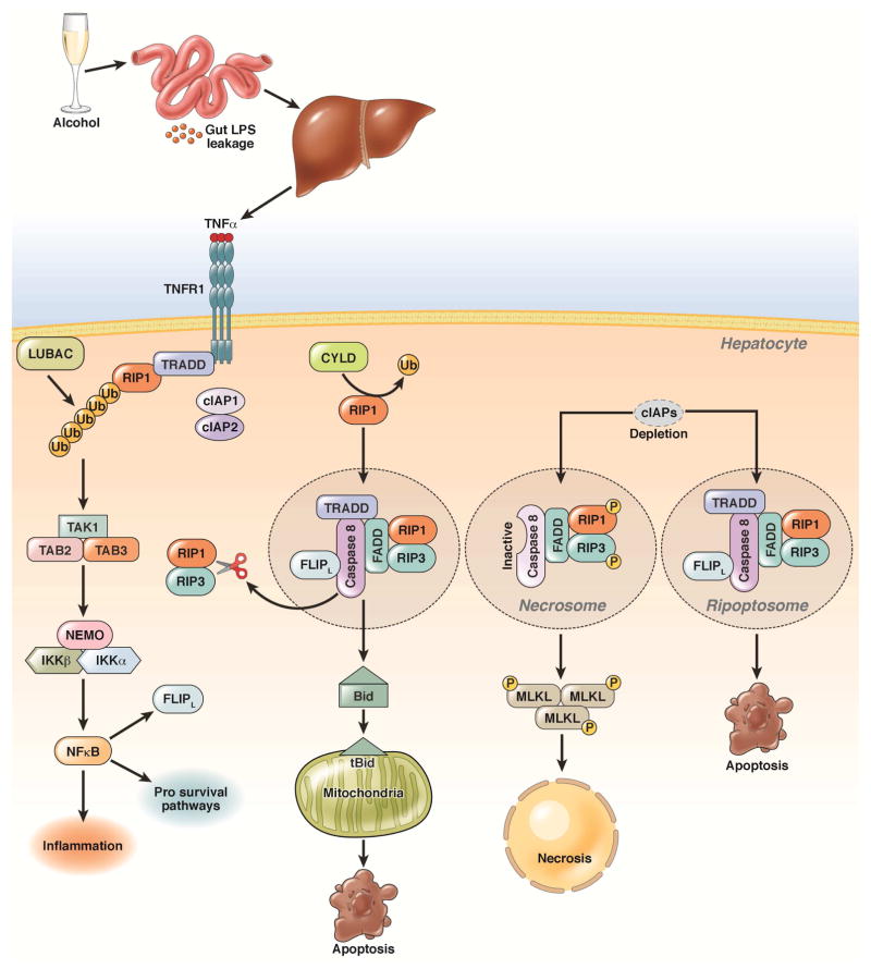
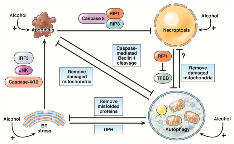
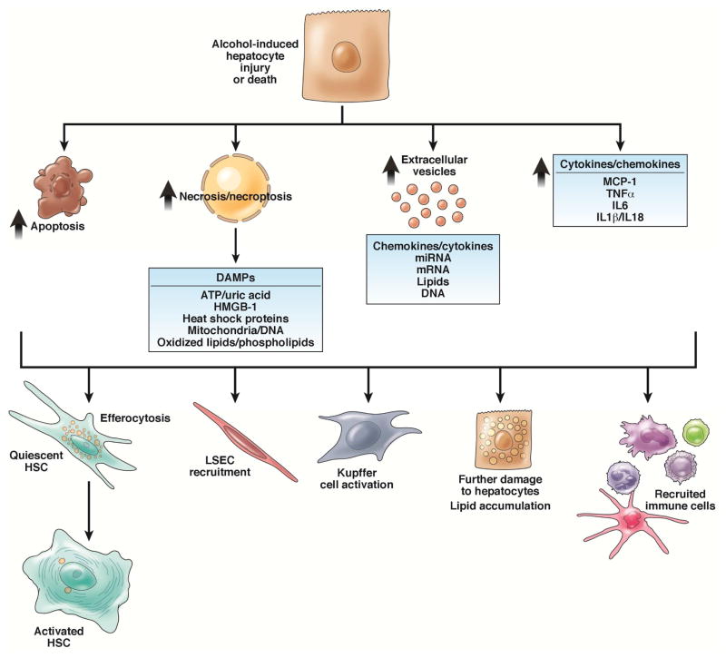
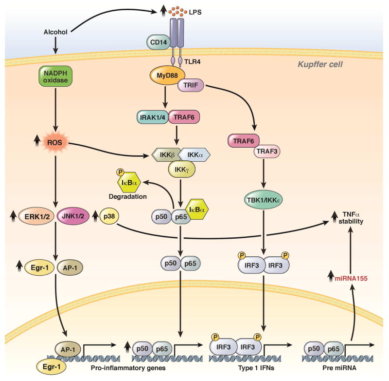
Alcoholic liver disease (ALD) develops in approximately 20% of alcoholic patients, with a higher prevalence in females. ALD progression is marked by fatty liver and hepatocyte necrosis, as well as apoptosis, inflammation, regenerating nodules, fibrosis, and cirrhosis.(1) ALD develops via a complex process involving parenchymal and nonparenchymal cells, as well as recruitment of other cell types to the liver in response to damage and inflammation. Hepatocytes are damaged by ethanol, via generation of reactive oxygen species and induction of endoplasmic reticulum stress and mitochondrial dysfunction. Hepatocyte cell death via apoptosis and necrosis are markers of ethanol-induced liver injury. We review the mechanisms by which alcohol injures hepatocytes and the response of hepatic sinusoidal cells to alcohol-induced injury. We also discuss how recent insights into the pathogenesis of ALD will affect the treatment and management of patients.

Keywords: Alcoholic Hepatitis; Alcoholic Liver Disease; Hepatic Stellate Cell.


Conflict of interest statement

DISCLOSURES: The authors disclose no conflicts.

Figures

Figure 1
Figure 1. Pathways of alcohol-induced apoptosis and necrosis
Alcohol consumption increases intestinal permeability resulting in increased influx of LPS into the liver. LPS activates Kupffer cells to produce TNF. TNF binds to its receptor (TNFR1), which recruits TNFR-associated death domain (TRADD), RIP1, cellular inhibitor of apoptosis proteins 1 (cIAP1/2), and linear ubiquitin chain assembly complex (LUBAC) and promotes the linear ubiquitination of RIP1. This leads to activation of NF-κB by the ubiquitin chain-dependent recruitment of transforming growth factor beta-activated kinase 1/MAP3K7 binding protein 2 (TAB2) and 3 (TAB3), transforming growth factor beta-activated kinase 1 (TAK1), and the I-κB kinase (IKK) complex. CYLD removes the ubiquitin chain from RIP1, and de-ubiquitinated RIP1 interacts with TRADD, fas-associated protein with a death domain (FADD), caspase-8, and the CASP8 and FADD-like apoptosis regulator (CFLAR), resulting in caspase-8 activation. Activated caspase-8 cleaves Bid to activate the mitochondrial apoptotic pathway and induce apoptosis; it also cleaves RIP1 and RIP3 to inactivate RIP1–RIP3-mediated necroptosis. When cIAPs are depleted and caspase-8 is inhibited, RIP1 and RIP3 interact via RHIM domains to from the amyloid-like necrosome. Auto- and trans-phosphorylated RIP1 and RIP3 then recruit downstream MLKL to initiate necroptosis. In the absence of cIAPs, RIP1, RIP3, TRADD, caspase-8, and CFLAR form a complex called the ripoptosome, which activates caspase-8 and apoptosis and requires RIP1 kinase activity.
Figure 2
Figure 2. Interactions among apoptosis, necrosis, ER stress, and autophagy
Apoptosis suppresses necroptosis via caspase-8 cleavage and inactivation of RIP1 and RIP3. Apoptosis also inhibits autophagy by caspase-mediated cleavage of Beclin 1. The necroptosis protein RIP1 negatively regulates TFEB to repress autophagy. ER stress induces apoptosis by activating caspase-12 and -4, JNK, and the interferon regulator factor 3 (IRF3) signaling pathway. ER stress can also induce autophagy via UPR signaling. Autophagy inhibits apoptosis and necrosis by removing damaged mitochondria. Autophagy also relieves ER stress by degrading misfolded proteins and protein aggregates.
Figure 3
Figure 3. Injured hepatocytes signal to cells within the hepatic sinusoid
Cells within the hepatic sinusoid respond to hepatocytes dying via apoptosis and necrosis/necroptosis, either via efferocytosis or phagocytosis of apoptotic bodies or DAMP-mediated signaling. Injured hepatocytes can also signal to their surrounding environment via the release of extracellular vesicles, containing a number of regulatory factors, or the direct expression of cytokines and chemokines.
Figure 4
Figure 4. Sensitization of TLR4-mediated signal transduction in hepatic macrophages
Chronic ethanol feeding not only increases the amount of circulating gut-derived endotoxin (LPS), but also sensitizes Kupffer cells to TLR4-mediated signaling via MyD88-dependent and –independent pathways. These processes increase expression of TLR4 and increase free radical formation by NADPH oxidase and CYP2E1. Increased ROS then increases TLR4-mediated signaling via MyD88-dependent activation of MAPKs and NF-κB. These signaling pathways integrate to increase transcription of cytokines, as well as the mRNA stability of TNF. This sensitization depends on ethanol-induced changes expression of microRNAs—particularly related to the stabilization of TNF mRNA. MyD8-independent signaling pathways activate STAT transcription factors and promote interferon production to regulate other immune cells in the liver.

References

    1. Crabb DW, Matsumoto M, Chang D, et al. Overview of the role of alcohol dehydrogenase and aldehyde dehydrogenase and their variants in the genesis of alcohol-related pathology. Proc Nutr Soc. 2004;63:49–63. - PubMed
    1. Lu Y, Cederbaum AI. CYP2E1 and oxidative liver injury by alcohol. Free Radic Biol Med. 2008;44:723–38. - PMC - PubMed
    1. Li Y, Wang S, Ni HM, et al. Autophagy in alcohol-induced multiorgan injury: mechanisms and potential therapeutic targets. Biomed Res Int. 2014;2014:498491. - PMC - PubMed
    1. Cao Q, Mak KM, Lieber CS. Cytochrome P4502E1 primes macrophages to increase TNF-alpha production in response to lipopolysaccharide. Am J Physiol Gastrointest Liver Physiol. 2005;289:G95–107. - PubMed
    1. Thakur V, Pritchard MT, McMullen MR, et al. Chronic ethanol feeding increases activation of NADPH oxidase by lipopolysaccharide in rat Kupffer cells: role of increased reactive oxygen in LPS-stimulated ERK1/2 activation and TNF-alpha production. J Leukoc Biol. 2006;79:1348–56. - PMC - PubMed