. 2016 Feb 26:13:48.
doi: 10.1186/s12974-016-0512-z.

Dual roles of the adenosine A2a receptor in autoimmune neuroinflammation

Affiliations

Dual roles of the adenosine A2a receptor in autoimmune neuroinflammation

J Ingwersen et al. J Neuroinflammation. .

Abstract

Background: Conditions of inflammatory tissue distress are associated with high extracellular levels of adenosine, due to increased adenosine triphosphate (ATP) degradation upon cellular stress or the release of extracellular ATP upon cell death, which can be degraded to adenosine by membrane-bound ecto-enzymes like CD39 and CD73. Adenosine is recognised to mediate anti-inflammatory effects via the adenosine A2a receptor (A2aR), as shown in experimental models of arthritis. Here, using pharmacological interventions and genetic inactivation, we investigated the roles of A2aR in experimental autoimmune encephalomyelitis (EAE), an animal model of multiple sclerosis (MS).

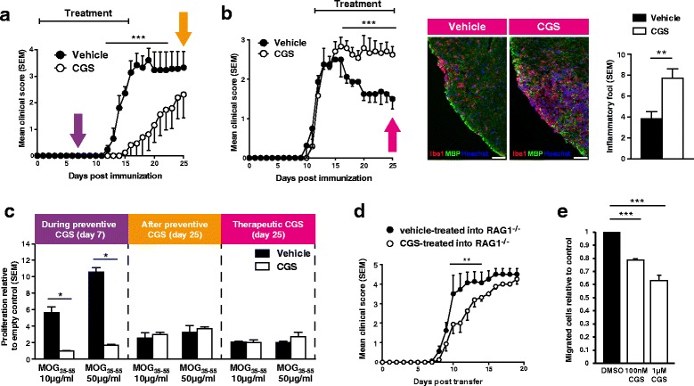
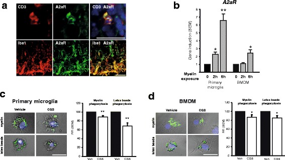
Methods: We used two independent mouse EAE variants, i.e. active immunization in C57BL/6 with myelin oligodendrocyte glycoprotein (MOG)35-55 or transfer-EAE by proteolipid protein (PLP)139-155-stimulated T lymphocytes and EAE in mice treated with A2aR-agonist CGS21680 at different stages of disease course and in mice lacking A2aR (A2aR(-/-)) compared to direct wild-type littermates. In EAE, we analysed myelin-specific proliferation and cytokine synthesis ex vivo, as well as inflammation and demyelination by immunohistochemistry. In vitro, we investigated the effect of A2aR on migration of CD4(+) T cells, macrophages and microglia, as well as the impact of A2aR on phagocytosis of macrophages and microglia. Statistical tests were Mann-Whitney U and Student's t test.

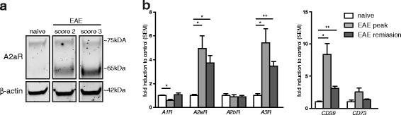
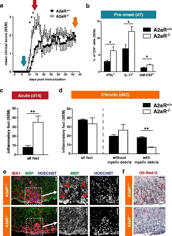
Results: We found an upregulation of A2aR in the central nervous system (CNS) in EAE, predominantly detected on T cells and macrophages/microglia within the inflamed tissue. Preventive EAE treatment with A2aR-specific agonist inhibited myelin-specific T cell proliferation ex vivo and ameliorated disease, while application of the same agonist after disease onset exacerbated non-remitting EAE progression and resulted in more severe tissue destruction. Accordingly, A2aR-deficient mice showed accelerated and exacerbated disease manifestation with increased frequencies of IFN-γ-, IL-17- and GM-CSF-producing CD4(+) T helper cells and higher numbers of inflammatory lesions in the early stage. However, EAE quickly ameliorated and myelin debris accumulation was lower in A2aR(-/-) mice. In vitro, activation of A2aR inhibited phagocytosis of myelin by macrophages and primary microglia as well as migration of CD4(+) T cells, macrophages and primary microglia.

Conclusions: A2aR activation exerts a complex pattern in chronic autoimmune neurodegeneration: while providing anti-inflammatory effects on T cells and thus protection at early stages, A2aR seems to play a detrimental role during later stages of disease and may thus contribute to sustained tissue damage within the inflamed CNS.


Figures

Fig. 1
Fig. 1
Expression of A2a receptors in autoimmune demyelination. a Expression of A2aR in naïve and active EAE (in C57BL/6 mouse strain) the spinal cord tissue. The protein A2aR has a molecular weight of approximately 42 kDA; the Western blotting band at ~65–70 kDa that we detected has been described for A2aR in the brain tissue before and is probably due to glycosylation [54, 55]. Use of a blocking peptide to the antibody against A2aR demonstrated specificity (see Additional file 1: Figure S1). b Expression of adenosine receptors and adenosine-generating ecto-enzymes in the spinal cord tissue in the course of adoptive transfer EAE (in SJL/J mouse strain). *p < 0.05, **p < 0.01
Fig. 2
Fig. 2
Effects of pharmacological A2aR activation in EAE. a Treatment with the A2aR-agonist CGS21680 (0.1 mg/kg) in the early phase of EAE leads to a prevention of disease symptoms (n = 8). Coloured arrows indicate the time points of ex vivo analyses. b Left: treatment with CGS21680 (0.1 mg/kg) in established disease (day 12 post immunization) leads to disease exacerbation (n = 5). Middle: histological analysis of CGS21680-treated EAE shows exacerbated Iba1+ accumulation in the spinal cord tissue (scale bars = 50 μm). Right: quantification of number of inflammatory foci at day 25 post immunization (n = 5). c Spleen cells isolated from vehicle-treated mice at day 7 in early-treatment paradigm (see purple arrow in Fig. 2a) show a higher antigen-specific proliferation capacity than cells isolated from CGS21680-treated mice. Cells isolated from late-stage early-treatment paradigm (orange arrow in Fig. 2a) and late-treatment paradigm (pink arrow in Fig. 2b) do not show differences in antigen-specific proliferation (n = 5). d T cells were transferred from EAE mice treated with CGS21680 or vehicle into naïve, non-immunised, non-treated RAG1-deficient mice (n = 4). e Analysis of migratory capacity towards CXCL12 in the presence or absence of CGS21680 of CD4+ T cells isolated from healthy mice using a Transwell chemotaxis system (data from three independent experiments). *p < 0.05, **p < 0.01, ***p < 0.001
Fig. 3
Fig. 3
EAE in A2aR-deficient mice. a EAE was induced in A2aR−/− and wild-type littermate C57BL/6 mice (n = 9). Coloured arrows indicate the time points of ex vivo analyses. b Ex vivo analysis of splenic CD4+ cells from A2aR−/− and wild-type animals at day 7 post immunization with MOG35-55 peptide (see blue arrow in Fig. 3a). Cells were cultured in the presence of the antigen and irradiated APC for 5 days and then analysed for their cytokine production by flow cytometry (n = 5). c Quantification of inflammatory foci in the spinal cord at acute phase of EAE (day 14, see red arrow in Fig. 3a). d Quantification of inflammatory foci in the spinal cord and association with myelin debris at chronic phase of EAE (day 42, see orange arrow in Fig. 3a). Bars on the left show all foci, bars on the middle and right show the same foci differentiated into foci with and without myelin debris. e Representative images of histological analysis of the spinal cord damage in chronic phase. The images on the right correspond to a magnification of the image on the left (indicated by dotted line and arrow). Red arrowheads indicate the area with disruption of myelin tissue. Scale bars = 50 μm. f Oil red O lipid stain visualisation of myelin debris in spinal cord lesions (day 42; see also Additional file 1: Figure S5). Scale bars = 100 μm. *p < 0.05, **p < 0.01
Fig. 4
Fig. 4
Analysis of A2aR-dependent modulation of phagocytosing cells. a CD3+ T cells and Iba1+ microglia/macrophages (red) co-stain for A2aR (green; in right overlay picture, yellow). Scale bars = 25 μm. b qPCR analysis of A2aR gene induction 2 and 6 h after addition of myelin to primary microglia and bone marrow-derived macrophages (BMDM) cultures. c, d Capacity of phagocytosis of labelled myelin lipoproteins and labelled carboxylate-modified polystyrene beads in primary microglia (c) and BMDM (d), treated with vehicle or CGS21680. Bar plots depict pooled data from 4–8 experiments. Representative images of microglia and macrophages with phagocytosed myelin and beads. Scale bars = 25 μm. *p < 0.05, **p < 0.01

References

    1. Linden J. Molecular approach to adenosine receptors: receptor-mediated mechanisms of tissue protection. Ann Rev Pharmacol Toxicol. 2001;41:775–787. doi: 10.1146/annurev.pharmtox.41.1.775. - DOI - PubMed
    1. Matzinger P. Tolerance, danger, and the extended family. Immunology. 1994;12:991–1045. doi: 10.1146/annurev.iy.12.040194.005015. - DOI - PubMed
    1. Kono H, Rock KL. How dying cells alert the immune system to danger. Nat Rev Immunol. 2008;8:279–289. doi: 10.1038/nri2215. - DOI - PMC - PubMed
    1. Blackburn MR, Vance CO, Morschl E, Wilson CN. Adenosine receptors and inflammation. Handb Exp Pharmacol. 2009;215–269. - PubMed
    1. Day Y, Huang L, McDuffie MJ, Rosin DL, Ye H, Chen J, et al. Renal protection from ischemia mediated by A2A adenosine receptors on bone marrow-derived cells. J Clin Invest. 2003;112:883–891. doi: 10.1172/JCI15483. - DOI - PMC - PubMed

MeSH terms

Substances

LinkOut - more resources