

Is the Lecompte technique the last word on transposition of the great arteries repair for all patients? A magnetic resonance imaging study including a spiral technique two decades postoperatively
- PMID: 26920722
- PMCID: PMC4986772
- DOI: 10.1093/icvts/ivw014
Is the Lecompte technique the last word on transposition of the great arteries repair for all patients? A magnetic resonance imaging study including a spiral technique two decades postoperatively
Abstract
Objectives: To compare the Lecompte technique and the spiral anastomosis (complete anatomic correction) two decades after arterial switch operation (ASO).
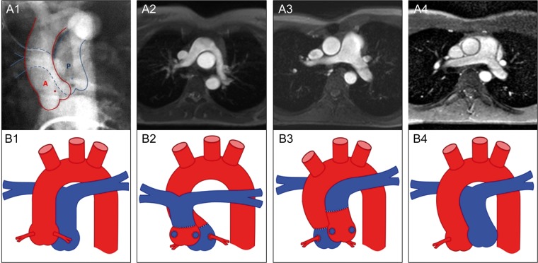
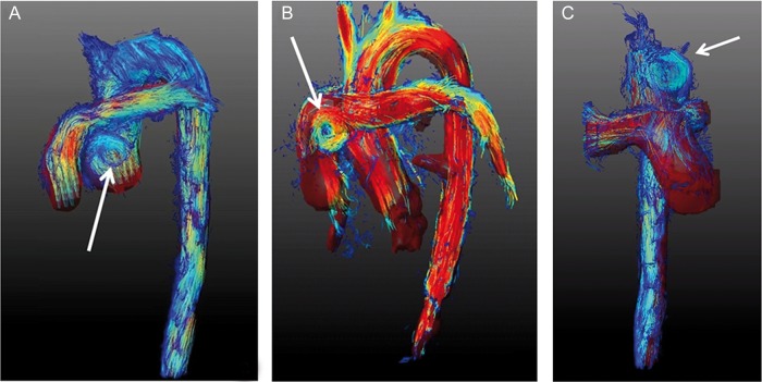
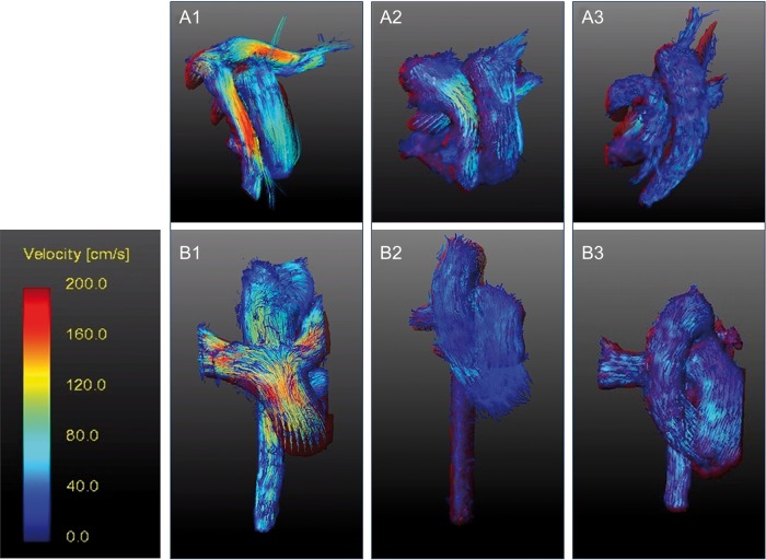
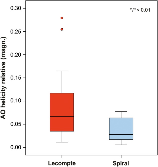
Methods: Nine patients after primary ASO with Lecompte and 6 selected patients after spiral anastomosis were evaluated 20.8 ± 2.1 years after ASO versus matched controls. Blood flow dynamics and flow profiles (e.g. vorticity, helicity) in the great arteries were quantified from time-resolved 3D magnetic resonance imaging (MRI) phase contrast flow measurements (4D flow MR) in addition to a comprehensive anatomical and functional cardiovascular MRI analysis.
Results: Compared with spiral reconstruction, patients with Lecompte showed more vortex formation, supranatural helical blood flow (relative helicity in aorta: 0.036 vs 0.089; P < 0.01), a reduced indexed cross-sectional area of the left pulmonary artery (155 vs 85 mm²/m²; P < 0.001) and more semilunar valve dysfunctions (n = 5 vs 1). There was no difference in elastic aortic wall properties, ventricular function, myocardial perfusion and myocardial fibrosis between the two groups. Cross-sectional area of the aortic sinus was larger in patients than in controls (669 vs 411 mm²/m²; P < 0.01). In the spiral group, the pulmonary root was rotated after ASO more towards the normal left position (P < 0.01).
Conclusions: In this study, selected patients with spiral anastomoses showed, two decades after ASO, better physiologically adapted blood flow dynamics, and attained a closer to normal anatomical position of their great arteries, as well as less valve dysfunction. Considering the limitations related to the small number of patients and the novel MRI imaging techniques, these data may provoke reconsidering the optimal surgical approaches to transposition of the great arteries repair.
Keywords: Lecompte technique; Magnetic resonance imaging; Physiological spiral anastomosis; Transposition of great vessels.
© The Author 2016. Published by Oxford University Press on behalf of the European Association for Cardio-Thoracic Surgery. All rights reserved.
Figures
References
-
- Liebman J, Cullum L, Belloc NB. Natural history of transposition of the great arteries. Anatomy and birth and death characteristics. Circulation 1969;40:237–62. - PubMed
-
- Jatene AD, Fontes VF, Paulista PP, de Souza LC, Neger F, Galantier M et al. Successful anatomic correction of transposition of the great vessels. A preliminary report. Arq Bras Cardiol 1975;28:461–4. - PubMed
-
- Lecompte Y, Zannini L, Hazan E, Jarreau MM, Bex JP, Tu TV et al. Anatomic correction of transposition of the great arteries. J Thorac Cardiovasc Surg 1981;82:629–31. - PubMed
-
- Lacour-Gayet F, Anderson RH. A uniform surgical technique for transfer of both simple and complex patterns of the coronary arteries during the arterial switch procedure. Cardiol Young 2005;15(Suppl 1):93–101. - PubMed
-
- Pretre R, Tamisier D, Bonhoeffer P, Mauriat P, Pouard P, Sidi D et al. Results of the arterial switch operation in neonates with transposed great arteries. Lancet 2001;357:1826–30. - PubMed
MeSH terms
LinkOut - more resources
Full Text Sources
Other Literature Sources
Medical
