. 2016 May;203(1):283-98.
doi: 10.1534/genetics.115.186270. Epub 2016 Feb 26.

Host Mitochondrial Association Evolved in the Human Parasite Toxoplasma gondii via Neofunctionalization of a Gene Duplicate

Affiliations

Host Mitochondrial Association Evolved in the Human Parasite Toxoplasma gondii via Neofunctionalization of a Gene Duplicate

Yaw Adomako-Ankomah et al. Genetics. 2016 May.

Abstract

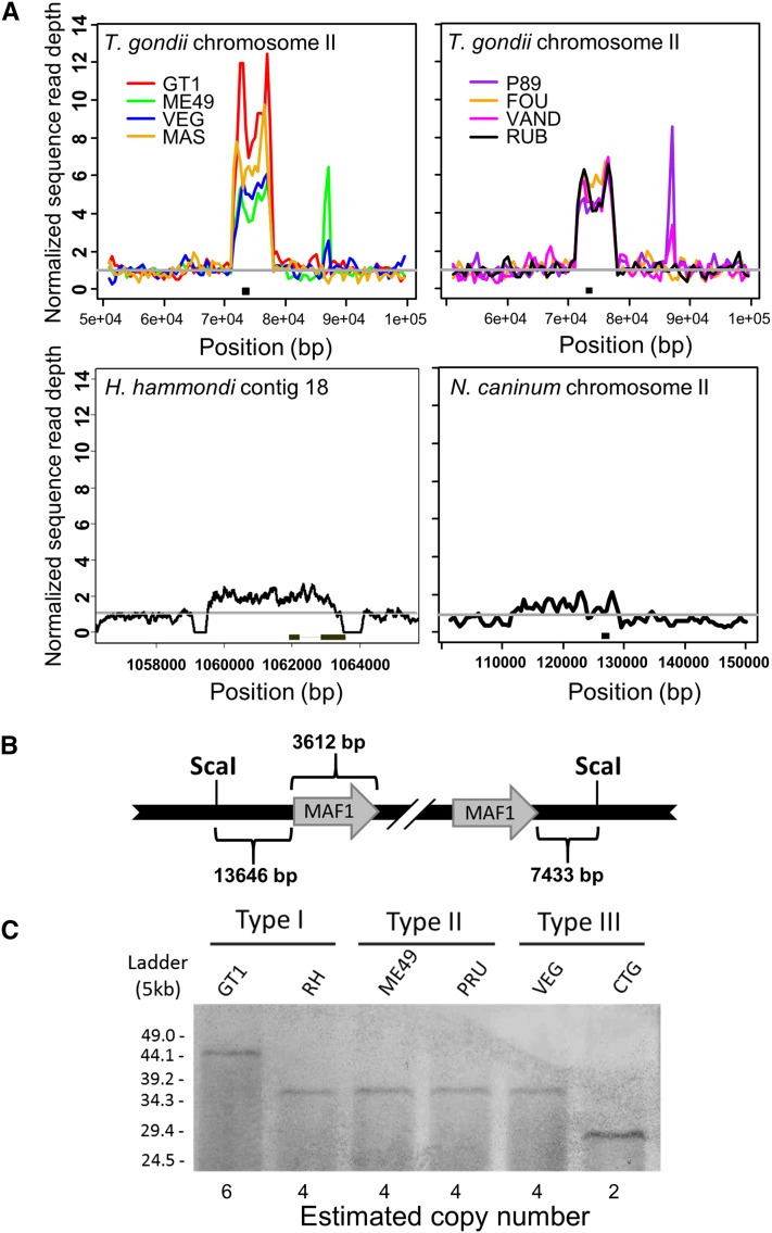
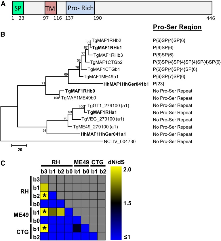
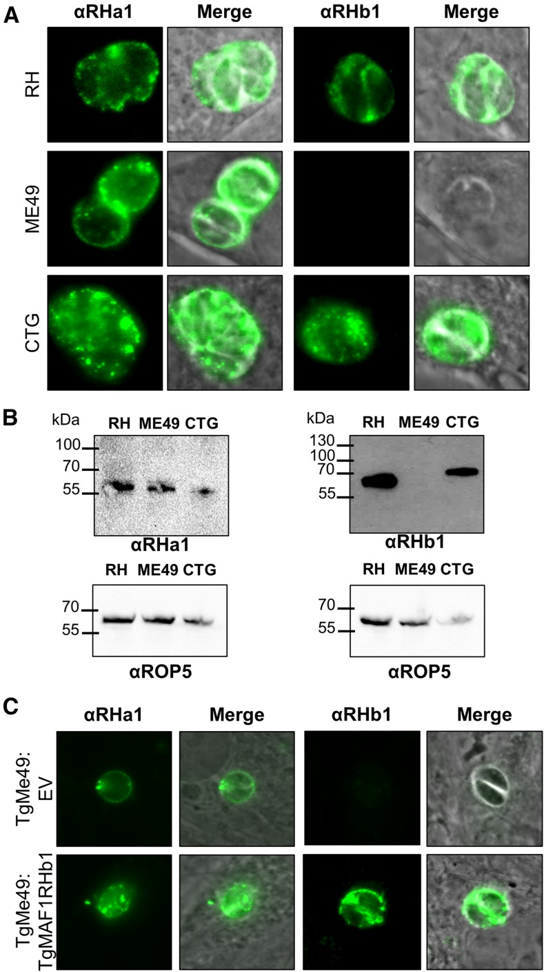
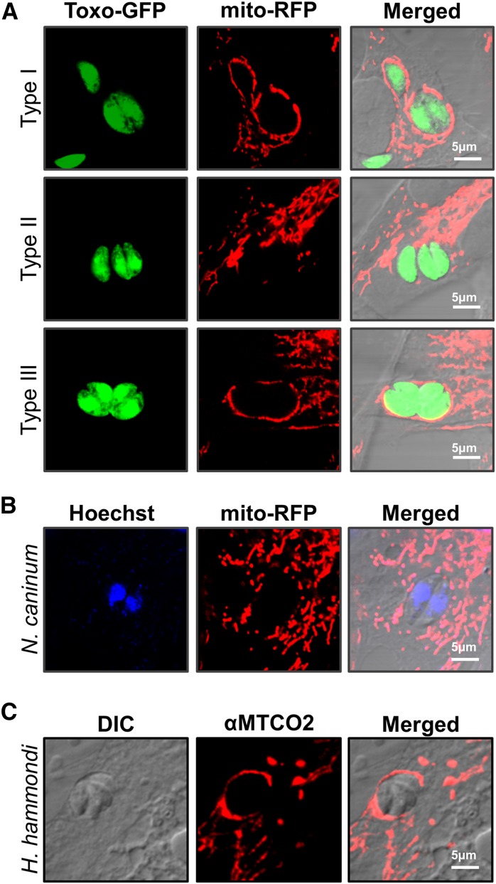
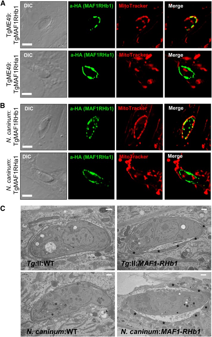
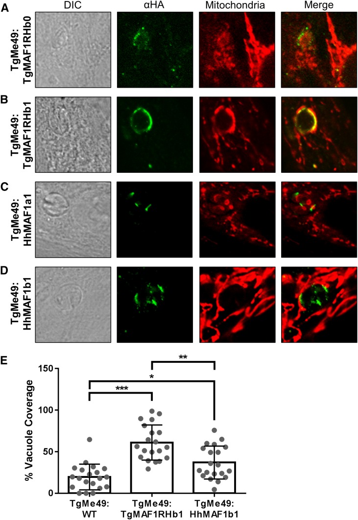
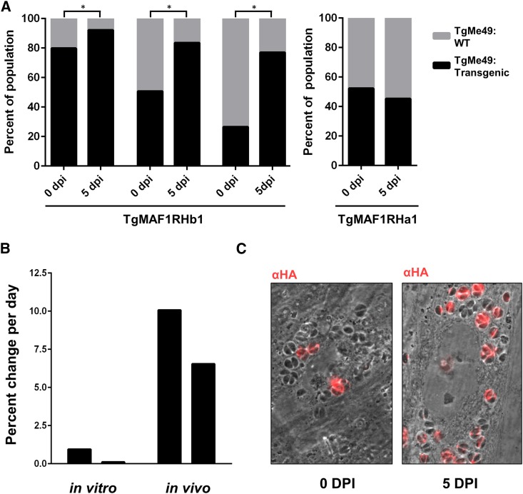
In Toxoplasma gondii, an intracellular parasite of humans and other animals, host mitochondrial association (HMA) is driven by a gene family that encodes multiple mitochondrial association factor 1 (MAF1) proteins. However, the importance of MAF1 gene duplication in the evolution of HMA is not understood, nor is the impact of HMA on parasite biology. Here we used within- and between-species comparative analysis to determine that the MAF1 locus is duplicated in T. gondii and its nearest extant relative Hammondia hammondi, but not another close relative, Neospora caninum Using cross-species complementation, we determined that the MAF1 locus harbors multiple distinct paralogs that differ in their ability to mediate HMA, and that only T. gondii and H. hammondi harbor HMA(+) paralogs. Additionally, we found that exogenous expression of an HMA(+) paralog in T. gondii strains that do not normally exhibit HMA provides a competitive advantage over their wild-type counterparts during a mouse infection. These data indicate that HMA likely evolved by neofunctionalization of a duplicate MAF1 copy in the common ancestor of T. gondii and H. hammondi, and that the neofunctionalized gene duplicate is selectively advantageous.

Keywords: Hammondia hammondi; Neospora caninum; Toxoplasma gondii; gene duplication; neofunctionalization.


Figures

Figure 1
Figure 1
The MAF1 locus exhibits copy number variation across strains of T. gondii and has comparatively low copy number in H. hammondi and N. caninum. (A) Coverage depth analysis for the MAF1 locus in eight T. gondii strain types and for the syntenic locus in H. hammondi and N. caninum. T. gondii sequences are from ToxoDB v7.3. Portions of the upper left panel of this figure were similarly represented in Pernas et al. (2014). Raw reads were plotted as described in Materials and Methods and normalized to the coverage 20 Kb upstream of the repetitive locus. Arrowheads indicate the location of predicted gene sequences based on ToxoDB (v7.3 for T. gondii; v26 for all other species). Asterisks indicate smaller repetitive sequence unrelated to MAF1 (see Materials and Methods for further explanation). (B) Schematic representation of the MAF1 locus showing ScaI restrictions sites outside of the locus, the size of the regions flanking the MAF1 locus, and the size of the repeat unit used to estimate copy number based on Southern blotting. The most relevant T. gondii ME49 gene name is indicated (from ToxoDB v7.3), although it does not fully match the sequenced paralogs. (C) ScaI-digested gDNA from each of six T. gondii strains was resolved by PFGE and probed with a MAF1-specific probe. The blot shows copy number variation consistent with predictions from sequence coverage analysis for strain types GT1, ME49, and VEG. Copy number for each strain was determined based on the schematic presented in B.
Figure 2
Figure 2
The T. gondii and H. hammondi MAF1 loci harbor two distinct isoforms while only one isoform is present in N. caninum. (A) Schematic representation of the predicted MAF1 protein. The signal peptide (SP) was predicted using SignalP v4.0 and the putative transmembrane domain (TM) was predicted by TMHMM v2.0. The proline-rich region (Pro-Rich) stretches from AA152 to 164 of TgMAF1RHb1 and is not found within all MAF1 paralogs (e.g., TgMAF1RHa1, a2). (B) Phylogram of either cloned MAF1 amino acid sequences from T. gondii, H. hammondi, and N. caninum, or those downloaded directly from ToxoDB (with TG Gene nos.). Cloned sequences of all of the “b” paralogs from T. gondii did not match any predicted gene models in ToxoDB in terms of predicted coding region length and were left out of the analysis. Paralog family is indicated at the end of each name (e.g., a1, b1, b2, etc.) (C) dN/dS ratio calculations for all T. gondii “b” MAF1 paralogs, including b0. * indicates significant evidence for diversifying selection for that particular paralog comparison (P < 0.05).
Figure 3
Figure 3
T. gondii MAF1 paralog expression differs between lineages. (A and B) Polyclonal antibodies were generated specifically against the C termini of TgMAF1RHa1 (Ser173 to Ser443) or TgMAF1RHb1 (Thr159 to Asp435). Protein expression was compared by immunofluorescence across three strains representing clonotypes I, II, and III. Based on immunofluorescence and Western blotting, antibodies against TgMAF1RHa1 detected protein in all three strains, while antibodies against TgMAF1RHb1 detected protein only in RH and CTG (and not ME49). (C) Antibodies against TgMAF1RHb1 are able to detect TgMAF1RHb1 expression in transgenic type II parasites expressing the TgMAF1RHb1 protein.
Figure 4
Figure 4
Host mitochondrial association is a feature of T. gondii and H. hammondi infections, but not N. caninum. (A) NRK-mitoRFP cells were infected with GFP-expressing type I, II, and III (RH, PRU, and CTG) parasites. Type II parasites are HMA, while types I and III are HMA+. (B) NRK-mitoRFP cells were infected with N. caninum strain NC-1. Cells were fixed and counterstained with Hoechst stain. Wild-type N. caninum are HMA. (C) HFFs were infected with H. hammondi sporozoites for 8 days before fixation. Host mitochondria were visualized using an antibody to human MTCO2. H. hammondi is HMA+.
Figure 5
Figure 5
MAF1RHa1 and MAF1RHb1 differ in their ability to complement HMA in T. gondii and N. caninum. (A) HFFs were labeled with MitoTracker and infected with parasites transiently transfected with either HA-MAF1RHa1 or HA-MAF1RHb1. MAF1RHb1 but not MAF1RHa1 is able to confer the HMA phenotype in TgME49. (B) Identical results were obtained for N. caninum. Bar, 5.0 μm. (C) HA-MAF1RHb1 was transfected into either TgME49 (top) or N. caninum (bottom), and HA-positive clones were isolated by limiting dilution. Wild type (WT, left) and TgMAF1RHb1 complemented (right) were grown for 18 hr in HFFs and processed for electron microscopy. Asterisks indicate host mitochondria. Bar, 500 nm.
Figure 6
Figure 6
N-terminally HA-tagged MAF1 isoforms were expressed in TgME49 parasites and HMA was assessed using MitoTracker or immunofluorescence assay using antibodies against the mitochondrial marker MTCO2. TgMAF1RHb0 and HhMAF1a1 (A and C) did not mediate HMA, while TgMAF1RHb1 and HhMAF1b1 (B and D) are both able to mediate HMA. (E) Quantification of percent vacuole coverage, determined by confocal microscopy. Twenty vacuoles were quantified for each of the MAF1 paralogs indicated, as well as wild-type TgME49. χ2 P-values: *0.0144; **0.0005; ***<0.0001.
Figure 7
Figure 7
Expression of TgMAF1RHb1, but not TgMAF1RHa1, in type II T. gondii increases competitive advantage. (A) Mice were infected with mixed populations of TgME49:EV and TgME49:MAF1 with the indicated isoforms and ratios. Infection was allowed to progress for 5 days and population proportions before and after infection were quantified by IFA. Both HMA+ and HMA MAF1 isoforms were assessed. TgME49:TgMAF1RHb1 significantly increases in proportion to TgME49:EV. *χ2 P-value <0.05. The proportion of TgMAF1RHa1-expressing parasites did not increase during the infection. (B) Percent change per day was calculated for the populations that started with 4:1 TgME49:EV to TgME49:TgMAF1RHb1 both in vitro and in vivo by dividing the total percent increase of TgMAF1RHb expressing parasites within the population by the number of days of infection. The first bar of both the in vitro and in vivo infections represent one clone set, while the second bar for each represents a second clone set. (C) Representative images of a mixed population from A before and after a 5-day in vivo infection. HA staining indicates TgMAF1RHb1-positive vacuoles.

References

    1. Adomako-Ankomah Y., Wier G. M., Borges A. L., Wand H. E., Boyle J. P., 2014. Differential locus expansion distinguishes Toxoplasmatinae species and closely related strains of Toxoplasma gondii. MBio 5: e01003–e01013. - PMC - PubMed
    1. Bahl A., Davis P. H., Behnke M., Dzierszinski F., Jagalur M., et al. , 2010. A novel multifunctional oligonucleotide microarray for Toxoplasma gondii. BMC Genomics 11: 603. - PMC - PubMed
    1. Bailey J. A., Eichler E. E., 2006. Primate segmental duplications: crucibles of evolution, diversity and disease. Nat. Rev. Genet. 7: 552–564. - PubMed
    1. Behnke M. S., Khan A., Wootton J. C., Dubey J. P., Tang K., et al. , 2011. Virulence differences in Toxoplasma mediated by amplification of a family of polymorphic pseudokinases. Proc. Natl. Acad. Sci. USA 108: 9631–9636. - PMC - PubMed
    1. Behnke M. S., Khan A., Lauron E. J., Jimah J. R., Wang Q., et al. , 2015. Rhoptry proteins ROP5 and ROP18 are major murine virulence factors in genetically divergent South American strains of Toxoplasma gondii. PLoS Genet. 11: e1005434. - PMC - PubMed

Publication types

Substances