. 2016 Feb 26;18(1):26.
doi: 10.1186/s13058-016-0686-4.

Mcl-1 confers protection of Her2-positive breast cancer cells to hypoxia: therapeutic implications

Affiliations

Mcl-1 confers protection of Her2-positive breast cancer cells to hypoxia: therapeutic implications

Muhammad Hasan Bashari et al. Breast Cancer Res. .

Erratum in

Abstract

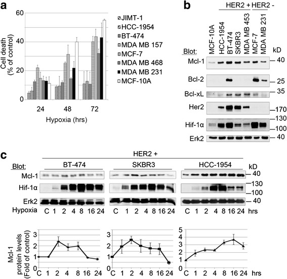
Background: Molecular mechanisms leading to the adaptation of breast cancer (BC) cells to hypoxia are largely unknown. The anti-apoptotic Bcl-2 family member myeloid cell leukemia-1 (Mcl-1) is frequently amplified in BC; and elevated Mcl-1 levels have been correlated with poor prognosis. Here we investigated the pathophysiologic role of Mcl-1 in Her2-positive BC cells under hypoxic conditions.

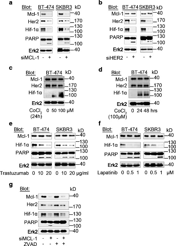
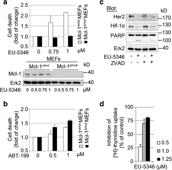
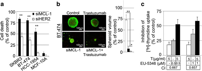
Methods: RNA interference and a novel small molecule inhibitor, EU-5346, were used to examine the role of Mcl-1 in Her2-positive BC cell lines and primary BC cells (sensitive or intrinsically resistant to Her2 inhibitors) under hypoxic conditions (using a hypoxic incubation chamber). Mechanisms-of-action were investigated by RT-PCR, mitochondrial isolation, as well as immunoprecipitation/blotting analysis, and microscopy. The specificity against Mcl-1 of the novel small molecule inhibitor EU5346 was verified in Mcl-1(Δ/null) versus Mcl-1(wt/wt) Murine Embryonic Fibroblasts (MEFs). Proliferation, survival, and spheroid formation were assessed in response to Mcl-1 and Her2 inhibition.

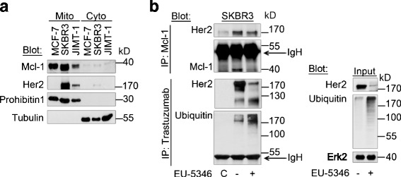
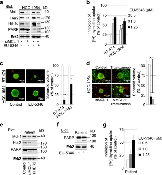
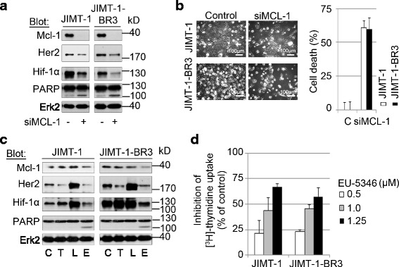
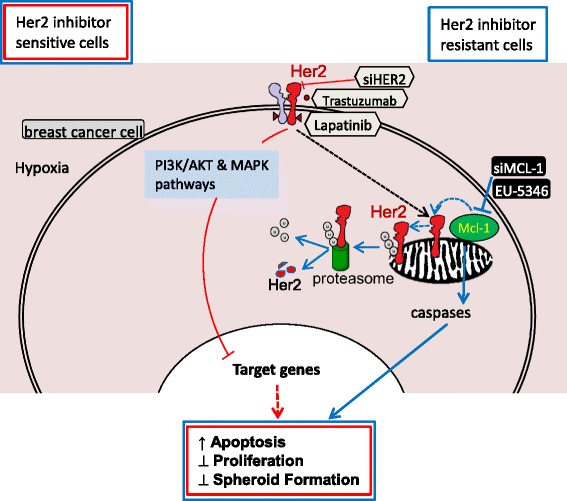
Results: We demonstrate for a strong correlation between high Mcl-1 protein levels and hypoxia, predominantly in Her2-positive BC cells. Surprisingly, genetic depletion of Mcl-1 decreased Her2 and Hif-1α levels followed by inhibition of BC cell survival. In contrast, Mcl-1 protein levels were not downregulated after genetic depletion of Her2 indicating a regulatory role of Mcl-1 upstream of Her2. Indeed, Mcl-1 and Her2 co-localize within the mitochondrial fraction and form a Mcl-1/Her2- protein complex. Similar to genetically targeting Mcl-1 the novel small molecule Mcl-1 inhibitor EU-5346 induced cell death and decreased spheroid formation in Her2-positive BC cells. Of interest, EU-5346 induced ubiquitination of Mcl-1- bound Her2 demonstrating a previously unknown role for Mcl-1 to stabilize Her2 protein levels. Importantly, targeting Mcl-1 was also active in Her2-positive BC cells resistant to Her2 inhibitors, including a brain-primed Her2-positive cell line.

Conclusion: Our data demonstrate a critical role of Mcl-1 in Her2-positive BC cell survival under hypoxic conditions and provide the preclinical framework for the therapeutic use of novel Mcl-1- targeting agents to improve patient outcome in BC.


Figures

Fig. 1
Fig. 1
Mcl-1 expression correlates with improved adaptation of Her2-positive BC cell lines to hypoxia. a Improved adaptation to hypoxic conditions of Her2-positive versus Her2-negative BC cells and the benign MCF-10A cell line. BC cell lines were incubated under hypoxic conditions for the time periods indicated. Cell survival was analyzed by MTT assay. Data represent mean ± SD for triplicate samples. Results shown are representative of three independent experiments. b Significant protein levels of Mcl-1, but not Bcl-2 or Bcl-xL, in BC cells under hypoxic conditions. BC cell lines or the nonmalignant MCF-10A cells were incubated for 4 hours under hypoxic conditions and whole-cell extracts were analyzed by immunoblotting with indicated antibodies. Immunoblotting for Erk2 confirmed equal protein loading. c Upregulation of Mcl-1 in Her2-positive BC cells under hypoxic conditions. BC cell lines were incubated under hypoxic conditions for up to 24 hours. Whole-cell extracts were analyzed by immunoblotting with indicated antibodies. Immunoblotting for Erk2 confirmed equal protein loading. Densitometric measurements (NIH ImageJ software) [59] were used to quantitate Mcl-1 protein levels from western blot images normalized for Erk2. Densitometric data represent mean values ± SD. Data shown are representative of three independent experiments. C normoxic control, Erk2 extracellular signal-regulated kinase 2, Her2 human epidermal growth factor receptor 2, Hif hypoxia-inducible factor, Mcl-1 myeloid cell leukemia-1
Fig. 2
Fig. 2
Mcl-1 is an upstream regulator of Her2 under hypoxic conditions. a Genetic depletion of Mcl-1 in Her2-positive BC cells promotes downregulation of Her2 and Hif-1α followed by inhibition of BC cell survival. b Mcl-1 protein levels do not decrease after genetically downregulating Her2. a, b BC cells were transfected with siMCL1 a or siHER2 b for 2 days and then exposed to hypoxia for 6 hours. c, d CoCl2-mediated stabilization of Hif-1α does not alter Mcl-1 or Her2 levels. BT-474 cells were exposed to CoCl2with indicated doses for 24 hours c and 100 μM CoCl2 for the indicated time periods d. e, f Pharmacologically targeting Her2 decreases Hif-1α but not Mcl-1 protein levels. BC cells were treated with the indicated doses of trastuzumab e or lapatinib f for 2 days and then exposed to hypoxia for 6 hours. g siMCL1-induced downregulation of Her2 and Hif-1α is not triggered by caspase-mediated off-target effects of MCL1 siRNA. Her2-positive BC cells were transfected with siMCL1 for 2 days and then exposed to hypoxia for 6 hours. ZVAD (50 μM) was added 24 hours before collecting samples. ag Whole-cell extracts were analyzed by immunoblotting with indicated antibodies. Immunoblotting for Erk2 confirmed equal protein loading. Erk2 extracellular signal-regulated kinase 2, Her2 human epidermal growth factor receptor 2, Hif hypoxia-inducible factor, kD kilodalton, Mcl-1 myeloid cell leukemia-1, PARP Poly (ADP-ribose) polymerase
Fig. 3
Fig. 3
The novel small molecule inhibitor EU-5346 specifically blocks Mcl-1 followed by cell death. a, b EU-5346 induces apoptosis in Mcl-1wt/wt but not Mcl-1Δ/null MEFs a. In contrast, ABT-199 induces apoptosis in both Mcl-1wt/wt and Mcl-1Δ/null MEFs b. MEFs were treated with indicated concentrations of EU-5346 a or ABT-199 b for 72 hours prior to Annexin V and PI staining. Data represent mean ± SD for triplicate samples. Results shown are representative of three independent experiments. c EU-5346-induced downregulation of Her2 and associated cell death are not triggered by caspase-mediated off-target effects of Mcl-1 siRNA. Her2-positive BC (SKBR3) cells were treated with EU-5346 and/or ZVAD (50 μM) and exposed to hypoxia during the last 6 hours. d EU-5346 inhibits proliferation of Her2-positive BC cells in a dose-dependent manner. BC cells were treated with EU-5346 for 3 days under hypoxic conditions. [3H]-thymidine was added during the last 8 hours. Data represent mean ± SD for triplicate samples. Results shown are representative of three independent experiments. c, d Whole-cell extracts were analyzed by immunoblotting with indicated antibodies. Immunoblotting for Erk2 confirmed equal protein loading. Erk2 extracellular signal-regulated kinase 2, Her2 human epidermal growth factor receptor 2, Hif hypoxia-inducible factor, kD kilodalton, Mcl-1 myeloid cell leukemia-1, MEF murine embryonic fibroblast, PARP Poly (ADP-ribose) polymerase
Fig. 4
Fig. 4
Her2 colocalizes with Mcl-1 within the mitochondrial fraction. a Mcl-1 and Her2 colocalize within the mitochondrial fraction. Mitochondrial and cytosolic proteins were isolated using the Qproteome Mitochondria Isolation Kit (Qiagen) according to the manufacturer’s instructions and analyzed by immunoblotting with indicated antibodies. Anti-Prohibitin 1 was used as a mitochondrial marker and anti-tubulin was used as a cytoplasmic marker. BC cells were exposed to hypoxia for 6 hours. b EU-5346 induces ubiquitination of Mcl-1-bound Her2. SKBR3 cells were treated with EU-5346 for 3 days and exposed to hypoxia during the last 6 hours. Whole-cell extracts were immunoprecipitated (IP) with either Mcl-1 antibody or trastuzumab and analyzed by immunoblotting with indicated antibodies. Nonspecific protein binding and detection were excluded by incubating protein A-Sepharose beads with lysis buffer and Mcl-1 antibody only (left panel). Input (right panel). C control, Erk2 extracellular signal-regulated kinase 2, Her2 human epidermal growth factor receptor 2, IgH heavy chain, kD kilodalton, Mcl-1 myeloid cell leukemia-1
Fig. 5
Fig. 5
Synergistic effects of combining trastuzumab with Mcl-1-targeting approaches in trastuzumab-sensitive Her2-positive BC cells. a Genetic depletion of Mcl-1 or Her2 induces cell death in Her2-positive BC cells, but not benign breast cells (MCF-10A). BC and MCF-10A cells were transfected with siMCL1 (filled bars) or siHER2 (open bars) for 30 hours and then exposed to hypoxia for 2 days. Cell survival was determined by AlamarBlue® assay. Data represent mean ± SD for triplicate samples. Results shown are representative of three independent experiments. b Synergistic effects of combining trastuzumab with siMCL1 in Her2-positive BC cells. BT-474 cells (5.5 × 103 cells) were seeded per well in agar-coated 96-well plates prior to siMCL1 transfection. Spheroid formation was assessed in siMCL1-treated and/or trastuzumab-treated and control cells using an inverted fluorescence light microscope. Photographs (10× magnification) of spheroid formation are representative of each group and three independent experiments (left). Spheroid volumes were calculated as described in Materials and methods. Data represent mean ± SD (right). c Synergistic growth inhibition of trastuzumab and EU-5346 in trastuzumab-sensitive Her2-positive BC cells under hypoxic conditions. BT-474 cells were pretreated with trastuzumab or dimethyl sulfoxide (DMSO) for 24 hours under normoxic conditions followed by EU-5346 treatment or DMSO for 72 hours under hypoxic conditions. [3H]-thymidine was added during the last 8 hours. Data represent mean ± SD for triplicate samples. Results shown are representative of three independent experiments. Trastuzumab (white bars), EU-5346 (gray bars), and drug combination (black bars). Combination index (CI) was calculated using the Combosyn software as described in Materials and methods. a, b *p <0.05 and **p <0.001 by Student’s t test
Fig. 6
Fig. 6
Genetically and pharmacologically targeting Mcl-1 induces cell death also in trastuzumab-resistant Her2-positive BC cells. a Both genetic depletion of Mcl-1 (siMCL1) and EU-5346 induces apoptosis in trastuzumab-resistant HCC-1954 BC cells. BC cells were transfected with siMCL1 (left) for 2 days or treated with EU-5346 (right) for 3 days and then exposed to hypoxia for 6 hours. b EU-5346 inhibits proliferation in Her2 inhibitor-sensitive and inhibitor-resistant BC cells in a dose-dependent manner. c EU-5346 inhibits spheroid formation in both trastuzumab-sensitive (BT-474) and trastuzumab-resistant (HCC-1954) BC cells. d Combining trastuzumab with siMCL1 in Her2 inhibitor-resistant Her2-positive BC cells does not induce synergistic effects. HCC-1954 cells (5.5 × 103 cells) were seeded per well in agar-coated 96-well plates prior to siMCL1 transfection. c, d Spheroid formation was assessed in EU-5346-treated or siMCL1-treated and/or trastuzumab-treated and control cells using an inverted fluorescence light microscope. Photographs of spheroid formation (10× magnification) are representative of each group and three independent experiments (left). Spheroid volumes were calculated as described in Materials and methods. Data represent mean ± SD (right). *p <0.05 and **p < 0.001 by Student’s t test. e, f siMCL1 e and EU-5346 f but not siHER2 e induces apoptosis in multidrug/Her2 inhibitor-resistant patient BC cells. BC cells were transfected with siMCL1 or siHER2 e for 2 days or treated with EU-5346 f for 3 days and then exposed to hypoxia for 6 hours. a, e, f Whole-cell extracts were analyzed by immunoblotting with indicated antibodies. Immunoblotting for Erk2 confirmed equal protein loading. g EU-5346 inhibits proliferation in multidrug/Her2 inhibitor-resistant patient BC cells in a dose-dependent manner. b, g BC cells were treated with EU-5346 for 3 days under hypoxic conditions. [3H]-thymidine was added during the last 8 hours. Data represent mean ± SD for triplicate samples. Results shown are representative of three independent experiments. Erk2 extracellular signal-regulated kinase 2, Her2 human epidermal growth factor receptor 2, Hif hypoxia-inducible factor, kD kilodalton, Mcl-1 myeloid cell leukemia-1, PARP Poly (ADP-ribose) polymerase
Fig. 7
Fig. 7
Genetically and pharmacologically targeting Mcl-1 induces cell death in brain-primed Her2 inhibitor-resistant Her2-positive BC cells. a, b Genetic depletion of Mcl-1 induces apoptosis in both maternal Her2 inhibitor-resistant Her2-positive JIMT-1 cells as well as in brain-primed JIMT-1 BR3 cells. a BC cells were transfected with siMCL1 for 2 days and exposed to hypoxia for 6 hours. Whole-cell extracts were analyzed by immunoblotting with indicated antibodies. Immunoblotting for Erk2 confirmed equal protein loading. b BC cells were transfected with siMCL1 for 30 hours and then exposed to hypoxia for 2 days. Cell survival was determined by AlamarBlue® assay. Data represent mean ± SD for triplicate samples. Results shown are representative of three independent experiments. c EU-5346 overcomes Her2 inhibitor resistance in brain-primed Her2-positive BC cells. BC cells were treated with indicated drugs for 3 days and then exposed to hypoxia during the last 6 hours. Whole-cell extracts were analyzed by immunoblotting with indicated antibodies. Immunoblotting for Erk2 confirmed equal protein loading. d EU-5346 inhibits proliferation of brain-primed Her2-positive BC cells in a dose-dependent manner. BC cells were treated with EU-5346 for 3 days under hypoxic conditions. [3H]-thymidine was added during the last 8 hours. Data represent mean ± SD for triplicate samples. Results shown are representative of three independent experiments. C control (dimethyl sulfoxide), E EU-5346, Erk2 extracellular signal-regulated kinase 2, Her2 human epidermal growth factor receptor 2, Hif hypoxia-inducible factor, kD kilodalton, L lapatinib, Mcl-1 myeloid cell leukemia-1, T trastuzumab
Fig. 8
Fig. 8
Summary: Mcl-1 confers protection of Her2-positive BC cells to hypoxia—therapeutic implications. Homodimerization of Her2 with Her2 or heterodimerization of Her2 with Her3 enhances phospho-inositol 3-kinase (PI3K)/AKT and RAS/mitogen-activated protein kinase (MAPK) pathways, which regulate BC cell proliferation, survival, and migration, as well as angiogenesis [60, 61]. The Her2 inhibitor trastuzumab [62] binds to the C-terminal portion of Her2. Another Her2 inhibitor, lapatinib [63], binds to the ATP binding site of Her2, but also Her1. Similar to siHER2, trastuzumab and lapatinib inhibit downstream signaling events, induce apoptosis, and inhibit proliferation of BC cells (red lines). Here we show a novel role for the antiapoptotic Bcl-2 family member Mcl-1 in Her2-positive BC cell adaptation to hypoxia. Specifically, our results show that Mcl-1 forms a protein complex with Her2 at the mitochondrial membrane and stabilizes Her2 by inhibiting its ubiquitination. Conversely, genetically (siMCL1) or pharmacologically (EU-5346) targeting Mcl-1 triggers ubiquitination and proteosomal degradation of Her2, thereby inducing apoptosis, and inhibiting proliferation and spheroid formation under hypoxic conditions. In addition, our results indicate the existence of a Mcl-1-dependent survival pathway in Her2-positive BC cells, which is independent of the Mcl-1–Her2 axis supporting the therapeutic benefit of combining Her2 (red lines) and Mcl-1 inhibitor (blue lines). Importantly, based on these findings, targeting Mcl-1 is also active in Her2-positive BC cells resistant to Her2 inhibitors, including a brain-primed Her2-positive cell line. Her human epidermal growth factor receptor, Mcl-1 myeloid cell leukemia-1. (Color figure online)

Similar articles

Cited by

References

    1. Sørlie T, Perou CM, Tibshirani R, Aas T, Geisler S, Johnsen H, et al. Gene expression patterns of breast carcinomas distinguish tumor subclasses with clinical implications. Proc Natl Acad Sci U S A. 2001;98:10869–74. doi: 10.1073/pnas.191367098. - DOI - PMC - PubMed
    1. Inwald EC, Klinkhammer-Schalke M, Hofstädter F, Zeman F, Koller M, Gerstenhauer M, et al. Ki-67 is a prognostic parameter in breast cancer patients: results of a large population-based cohort of a cancer registry. Breast Cancer Res Treat. 2013;139:539–52. doi: 10.1007/s10549-013-2560-8. - DOI - PMC - PubMed
    1. Ellis MJ, Perou CM. The genomic landscape of breast cancer as a therapeutic roadmap. Cancer Discov. 2013;3:27–34. doi: 10.1158/2159-8290.CD-12-0462. - DOI - PMC - PubMed
    1. Perou CM, Sørlie T, Eisen MB, van de Rijn M, Jeffrey SS, Rees CA, et al. Molecular portraits of human breast tumours. Nature. 2000;406:747–52. doi: 10.1038/35021093. - DOI - PubMed
    1. Network CGA. Comprehensive molecular portraits of human breast tumours. Nature. 2012;490:61–70. doi: 10.1038/nature11453. - DOI - PMC - PubMed

Publication types

Substances