. 2016 Apr 29;291(18):9678-89.
doi: 10.1074/jbc.M116.720573. Epub 2016 Feb 26.

Co-fibrillogenesis of Wild-type and D76N β2-Microglobulin: THE CRUCIAL ROLE OF FIBRILLAR SEEDS

Affiliations

Co-fibrillogenesis of Wild-type and D76N β2-Microglobulin: THE CRUCIAL ROLE OF FIBRILLAR SEEDS

Antonino Natalello et al. J Biol Chem. .

Abstract

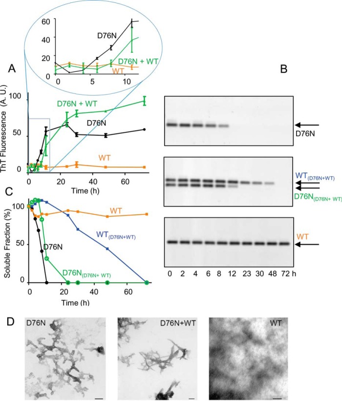
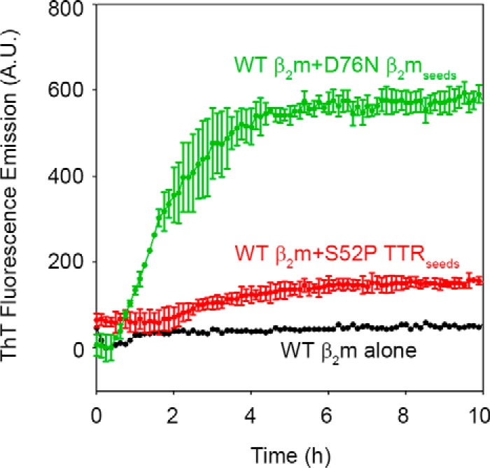
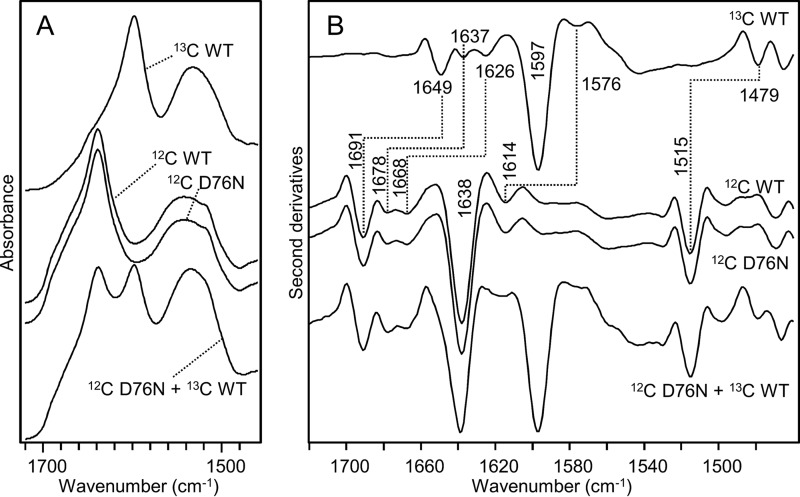
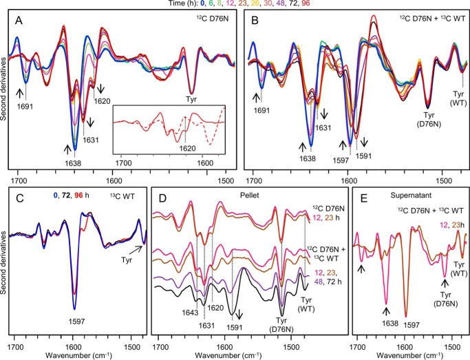
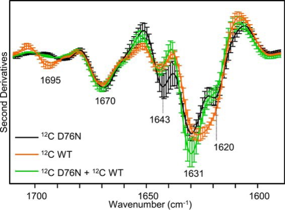
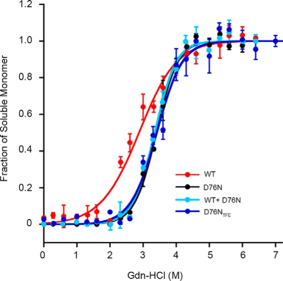
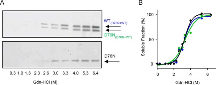
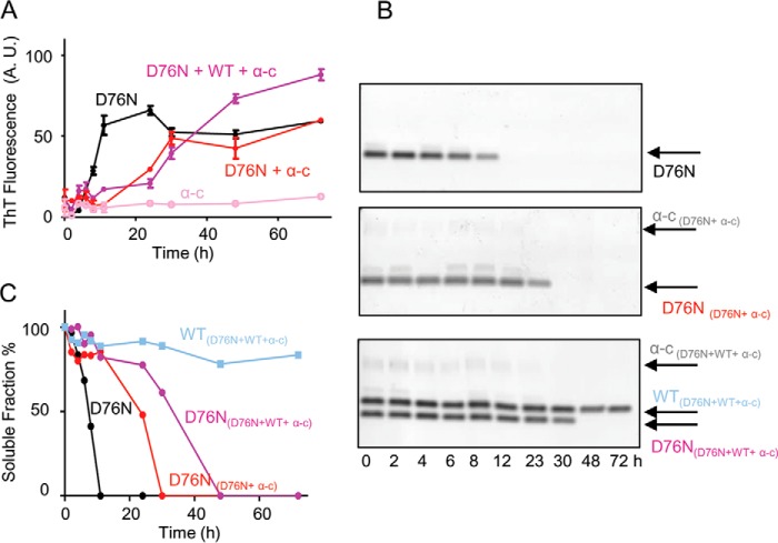
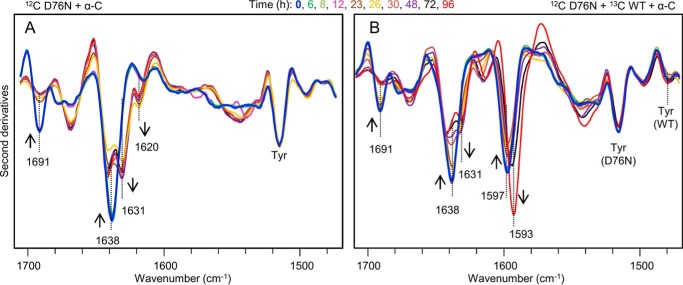
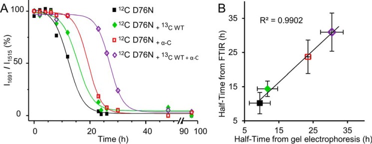
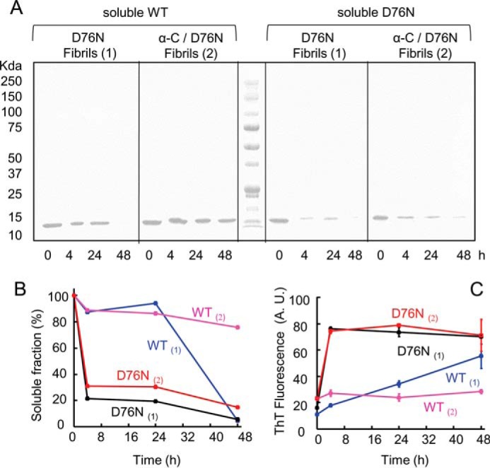
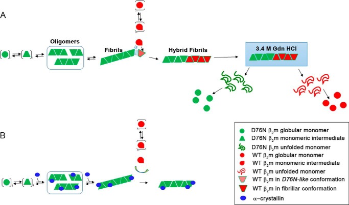
The amyloidogenic variant of β2-microglobulin, D76N, can readily convert into genuine fibrils under physiological conditions and primes in vitro the fibrillogenesis of the wild-type β2-microglobulin. By Fourier transformed infrared spectroscopy, we have demonstrated that the amyloid transformation of wild-type β2-microglobulin can be induced by the variant only after its complete fibrillar conversion. Our current findings are consistent with preliminary data in which we have shown a seeding effect of fibrils formed from D76N or the natural truncated form of β2-microglobulin lacking the first six N-terminal residues. Interestingly, the hybrid wild-type/variant fibrillar material acquired a thermodynamic stability similar to that of homogenous D76N β2-microglobulin fibrils and significantly higher than the wild-type homogeneous fibrils prepared at neutral pH in the presence of 20% trifluoroethanol. These results suggest that the surface of D76N β2-microglobulin fibrils can favor the transition of the wild-type protein into an amyloid conformation leading to a rapid integration into fibrils. The chaperone crystallin, which is a mild modulator of the lag phase of the variant fibrillogenesis, potently inhibits fibril elongation of the wild-type even once it is absorbed on D76N β2-microglobulin fibrils.

Keywords: Fourier transform IR (FTIR); amyloid; fibril; protein aggregation; protein misfolding; β2-microglobulin.


Figures

FIGURE 1.
FIGURE 1.
Fibrillogenesis of D76N and WT β2m. A, the time course of aggregation of D76N β2m, WT β2m, equimolar mixture of D76N β2m and WT β2m under stirring conditions, at 37 °C, was monitored by ThT fluorescence emission with excitation and emission wavelengths at 445 and 480 nm, respectively. Inset, expanded view of the ThT signal between 0 and 12 h. B, agarose gel electrophoresis analysis of supernatants from fibrillogenesis samples as described above. The arrows show the electrophoretic mobility of each isoform. C, density of agarose gel bands were measured and plotted as soluble fractions with time. Values shown in A and C are mean ± S.D. (error bars) from three independent experiments. D, negatively stained transmission electron microscopy (scale bar, 100 nm) showing that only WT β2m alone does not form fibrils under physiological conditions and in the absence of D76N β2m seeds.
FIGURE 2.
FIGURE 2.
Cross-seeding fibrillogenesis of WT β2m. Time course of aggregation of WT β2m (40 μm) in the absence or presence of D76N β2m seeds (1.7 μm) or S52P TTR seeds (1.4 μm) as described under “Experimental Procedures.” Relative intensities of ThT emission, after subtraction of the corresponding seeds fluorescence, were plotted with time. Mean ± S.D. (error bars) from three independent experiments. A.U., arbitrary units.
FIGURE 3.
FIGURE 3.
FTIR spectra of unlabeled (12C) and isotopically labeled (13C) β2m. A, absorption spectra of native [12C]WT β2m, [13C]WT β2m, [12C]D76N, and an equimolar mixture of [13C]WT and [12C]D76N. B, second derivatives of absorption spectra shown in A. Peak positions of the main components are indicated. The absence of the peak at 1691 cm−1 in the 13C protein confirms that isotopic labeling was successfully achieved.
FIGURE 4.
FIGURE 4.
Time course of β2m aggregation studied by isotope-edited FTIR spectroscopy. A, second derivatives of absorption spectra of [12C]D76N variant β2m at 50 μm at different times of incubation at 37 °C (blue, time 0; red, time 96 h). Inset, expanded view of the second derivative spectra of the aggregates by [12C]D76N variant alone (solid line) and by [12C]D76N/[13C]WT β2m equimolar mixture (dashed line) after 96 h of incubation. B, second derivatives of absorption spectra of an equimolar mixture of [12C]D76N/[13C]WT β2m, both at 50 μm, at different times of incubation at 37 °C. C, second derivatives of absorption spectra of [13C]WT β2m at 50 μm at different times of incubation at 37 °C. D, second derivatives of absorption spectra of the pellet, and E, of the supernatant obtained by centrifugation of aliquots withdrawn from the same samples analyzed. Only spectra at selected incubation times are shown. The arrows point to the spectral changes occurring with time. Spectra are reported after normalization at the Tyr peak around 1515 cm−1 in D76N β2m (A, B, and D), at the Tyr peak of WT β2m (C), or at the native β-sheet peak at ∼1597 cm−1 in WT β2m (E).
FIGURE 5.
FIGURE 5.
Second derivative spectra of mature β2m fibrils. Second derivative spectra of fibrils by D76N β2m alone or by an equimolar mixture of WT and D76N β2m formed after incubation at 37 °C under shear forces and, by WT β2m at neutral pH in the presence of 20% TFE. Isotopically unlabeled proteins were used. Mean ± S.D. (Error bars) of spectra from 3 independent fibril preparations are shown.
FIGURE 6.
FIGURE 6.
Thermodynamic stability of in vitro fibrils. The proportion of monomer released from β2m fibrils over the total protein concentration at increasing GdnHCl concentrations was analyzed with Equation 1 following the linear polymerization model as described under “Experimental Procedures.”
FIGURE 7.
FIGURE 7.
WT and D76N β2m are simultaneously released from the hybrid fibrils. A, agarose gel electrophoresis analysis of refolded soluble fractions of fibrils formed under shear forces by an equimolar mixture of WT/D76N β2m or D76N β2m alone (see “Experimental Procedures”) at different denaturant concentrations. B, soluble fraction measured by density of gel bands was plotted with denaturant concentration showing that the same amount of WT and D76N β2m was released from the mixed fibrils and that a similar quantity of soluble D76N β2m was generated during the disassembly of the corresponding homogenous fibrils.
FIGURE 8.
FIGURE 8.
Fibrillogenesis of D76N β2m in the presence of α-crystallin and WT β2m. A, time course of aggregation of D76N β2m alone, D76N β2m in the presence of α-crystallin (α-C), an equimolar mixture of WT and D76N β2m in the presence of α-crystallin and, α-crystallin alone was monitored under stirring conditions by fluorescence emission of ThT. Protein concentrations were 50 μm for each β2m isoform and 10 μm for α-crystallin, respectively. Data are mean ± S.D. of three independent experiments. A.U., arbitrary units. B, agarose gel electrophoresis analysis of supernatants from one series of fibrillogenesis samples containing D76N β2m alone, D76N β2m in the presence of 10 μm α-crystallin, and equimolar mixture of WT and D76N β2m in the presence of 10 μm α-crystallin. C, soluble fraction quantified by density of gel bands and plotted with time.
FIGURE 9.
FIGURE 9.
Time course of β2m aggregation in the presence of α-crystallin studied by isotope-edited FTIR spectroscopy. A, second derivatives of absorption spectra of 50 μm [12C]D76N in the presence of 10 μm α-crystallin collected at different times of incubation, 37 °C. B, second derivatives of absorption spectra of an equimolar mixture of the two β2m species in the presence of α-crystallin (50 μm [12C]D76, 50 μm [13C]WT, 10 μm α-crystallin) at different times of incubation, 37 °C.
FIGURE 10.
FIGURE 10.
Time course of D76N aggregation. A, time course of the intensity of the ∼1691 cm−1 component of the D76N variant, due to the native β-sheet structures. The intensities at ∼1691 cm−1 were normalized at the tyrosine peak of the variant (at ∼1515 cm−1) and given as percentage variation. B, the aggregation half-time of D76N β2m under different conditions was obtained from the FTIR data and compared with that obtained from the electrophoretic analyses.
FIGURE 11.
FIGURE 11.
Residual soluble WT β2m or D76N β2m during aggregation in the presence of pre-formed fibrils. A, SDS-PAGE electrophoresis analysis of the soluble fraction of WT and D76N β2m at different times of incubation in PBS, 37 °C, under stirring conditions in the presence of D76N β2m fibrils alone (1) or in association with α-crystallin (2). B, density of gel bands in A were measured and plotted as soluble fractions with time. C, aggregation was monitored by ThT fluorescence emission with excitation and emission wavelengths at 445 and 480 nm, respectively.
FIGURE 12.
FIGURE 12.
AFM analysis of fibrils and interaction with α-crystallin. Surface plots of topographic AFM images showing fibrillar aggregates formed by D76N β2m alone and by the equimolar mixture of WT and D76N β2m, in the absence (top) and presence (bottom) of α-crystallin (α-cry). Globular structures can be also observed in the upper corner of the image of fibrils by D76N+α-cry or in the background of the image of fibrils by D76N+WT+α-cry.
FIGURE 13.
FIGURE 13.
Schematic representation of the mechanism of copolymerization of D76N and WT β2m. A, nucleation phase only involves native globular D76N β2m. When D76N fibrils are formed, the WT protein can start the fibrils elongation. Surface of the edge of fibrils facilitates the fibrillary conversion of monomeric WT β2m. Disassembly of hybrid WT/D76N β2m fibrils by chemical denaturation occurs via simultaneous release of WT and variant. B, crystallin absorbed on D76N β2m fibrils prevents their seeding effect on wild-type β2m, which, at this state, cannot contribute to fibril elongation.

Similar articles

Cited by

References

    1. Bellotti V., and Chiti F. (2008) Amyloidogenesis in its biological environment: challenging a fundamental issue in protein misfolding diseases. Curr. Opin. Struct. Biol. 18, 771–779 - PubMed
    1. Booth D. R., Sunde M., Bellotti V., Robinson C. V., Hutchinson W. L., Fraser P. E., Hawkins P. N., Dobson C. M., Radford S. E., Blake C. C., and Pepys M. B. (1997) Instability, unfolding and aggregation of human lysozyme variants underlying amyloid fibrillogenesis. Nature 385, 787–793 - PubMed
    1. Colon W., and Kelly J. W. (1992) Partial denaturation of transthyretin is sufficient for amyloid fibril formation in vitro. Biochemistry 31, 8654–8660 - PubMed
    1. Naiki H., Hashimoto N., Suzuki S., Kimura H., Nakakuki K., and Gejyo F. (1997) Establishment of a kinetic model of dialysis-related amyloid fibril extension in vitro. Amyloid 4, 223–232
    1. Valleix S., Gillmore J. D., Bridoux F., Mangione P. P., Dogan A., Nedelec B., Boimard M., Touchard G., Goujon J. M., Lacombe C., Lozeron P., Adams D., Lacroix C., Maisonobe T., Planté-Bordeneuve V., et al. (2012) Hereditary systemic amyloidosis due to Asp76Asn variant β2-microglobulin. N. Engl. J. Med. 366, 2276–2283 - PubMed

Publication types

LinkOut - more resources