. 2016 May 15;76(10):3045-56.
doi: 10.1158/0008-5472.CAN-15-2310. Epub 2016 Feb 26.

Extracellular Adenosine Production by ecto-5'-Nucleotidase (CD73) Enhances Radiation-Induced Lung Fibrosis

Affiliations

Extracellular Adenosine Production by ecto-5'-Nucleotidase (CD73) Enhances Radiation-Induced Lung Fibrosis

Florian Wirsdörfer et al. Cancer Res. .

Abstract

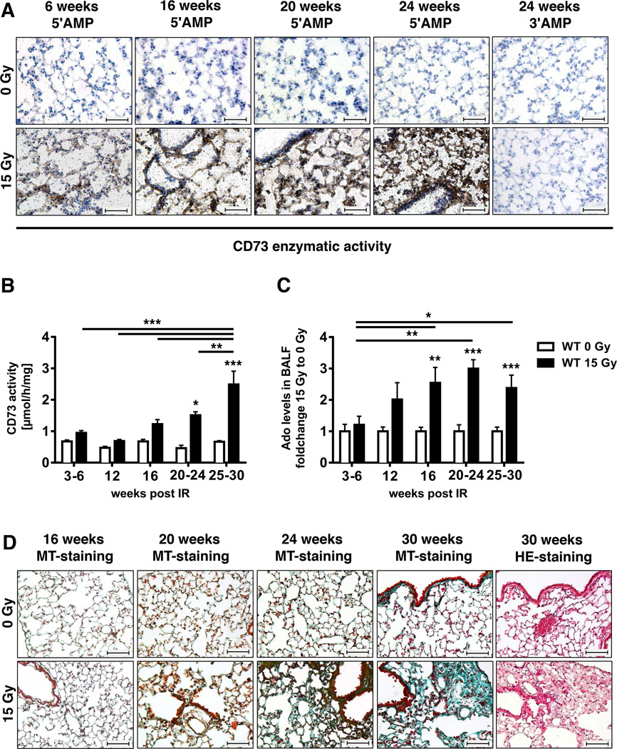
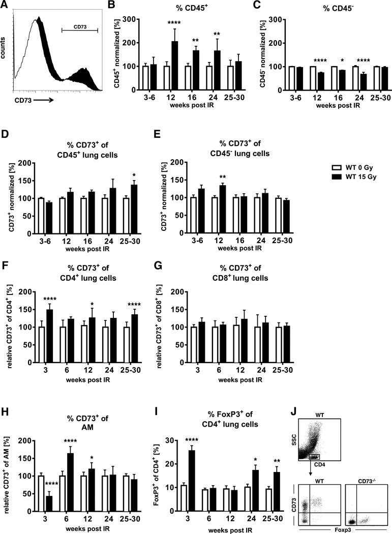
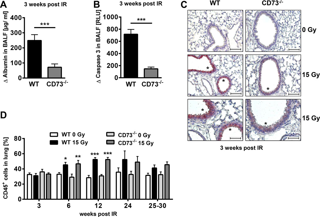
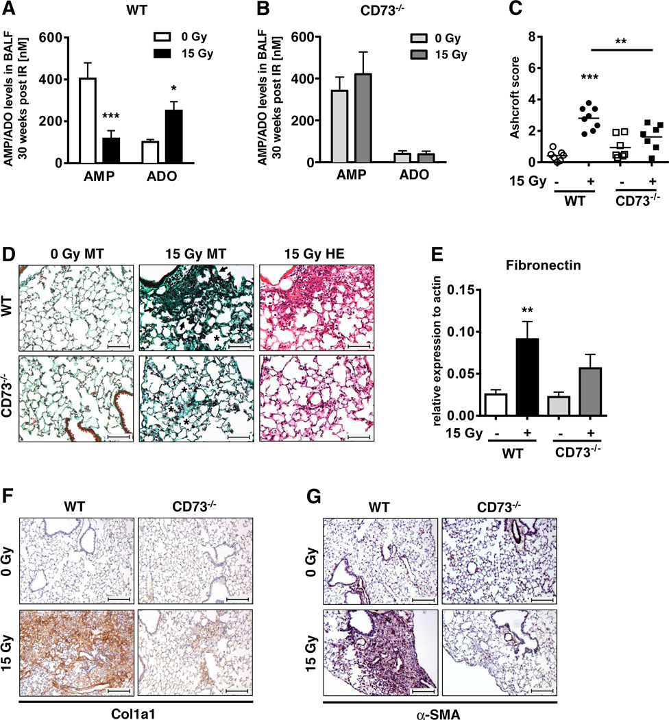
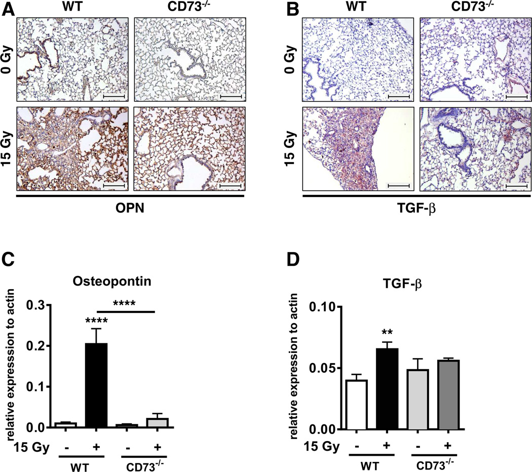
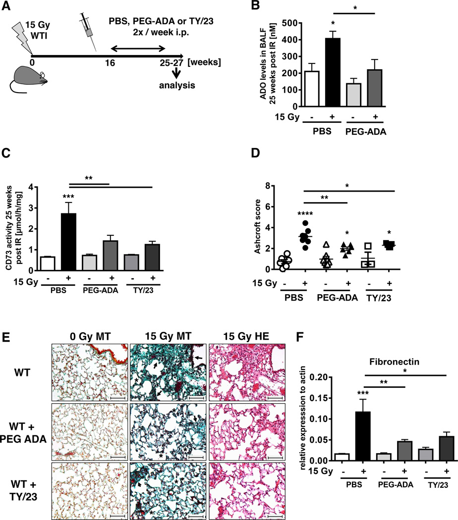
Radiation-induced pulmonary fibrosis is a severe side effect of thoracic irradiation, but its pathogenesis remains poorly understood and no effective treatment is available. In this study, we investigated the role of the extracellular adenosine as generated by the ecto-5'-nucleotidase CD73 in fibrosis development after thoracic irradiation. Exposure of wild-type C57BL/6 mice to a single dose (15 Gray) of whole thorax irradiation triggered a progressive increase in CD73 activity in the lung between 3 and 30 weeks postirradiation. In parallel, adenosine levels in bronchoalveolar lavage fluid (BALF) were increased by approximately 3-fold. Histologic evidence of lung fibrosis was observed by 25 weeks after irradiation. Conversely, CD73-deficient mice failed to accumulate adenosine in BALF and exhibited significantly less radiation-induced lung fibrosis (P < 0.010). Furthermore, treatment of wild-type mice with pegylated adenosine deaminase or CD73 antibodies also significantly reduced radiation-induced lung fibrosis. Taken together, our findings demonstrate that CD73 potentiates radiation-induced lung fibrosis, suggesting that existing pharmacologic strategies for modulating adenosine may be effective in limiting lung toxicities associated with the treatment of thoracic malignancies. Cancer Res; 76(10); 3045-56. ©2016 AACR.


Conflict of interest statement

Conflict of interest notification:

No conflict of interest does exist for any of the authors

Figures

Figure 1
Figure 1. Radiation induces up-regulation of CD73, adenosine accumulation and lung fibrosis
C57BL/6 WT mice received 0 or 15 Gy WTI and were sacrificed at the indicated time points post-irradiation. (A) CD73 histochemistry on lung frozen sections of irradiated and control mice using 5'-AMP or 5'-AMP (negative control) as substrates. CD73 activity is visualized by the black lead sulfide precipitate. (B) Lung CD73 enzyme activity at the indicated time points (n = 12/12, 9/9, 10/9, 5/4, 19/13 [0 Gy/15 Gy]). (C) Fold change of BALF adenosine levels (mean ± SEM) of irradiated vs. control mice. Means of control values were between 41 and 149.5 nM (n = 9/10, 7/7, 10/9, 8/7, 20/11 [0 Gy/15 Gy]). *P ≤ 0.05, **P ≤ 0.01, ***P ≤ 0.001 by two-way ANOVA followed by post-hoc Bonferroni test. (D) Masson´s Goldner Trichrome (MT) and H&E staining of paraffin-embedded lung sections (scale bar = 100 µm).
Figure 2
Figure 2. Thoracic irradiation triggers time-dependent alterations in CD73 expression on resident cells and immune cells
C57BL/6 WT mice received 0 or 15 Gy WTI and were sacrificed at the indicated time points post-irradiation. Whole lung cell (WLC) CD45 and/or CD73 expression were analyzed by flow cytometry. (A) CD73 expression in WLC is shown in the black histogram. (B, C) Normalized percentages of CD45+ (B, n = 11/11, 16/16, 6/6, 6/5, 10/12) or CD45 (C, n = 15/11, 16/16, 6/6, 6/5, 10/12) WLC. (D-H) Normalized percentages of CD45+, CD45, CD4+, CD8+ cells and alveolar macrophages (CD11chi autofluorescent (55)) with CD73 surface expression (D, n = 11/11, 16/16, 6/6, 6/5, 10/12; E, n = 11/11, 16/16, 6/6, 6/5, 9/11; F, G n = 7/7, 7/7, 14/14, 6/6, 15/15; H, n = 10/10, 8/8, 12/12, 6/4, 6/4). (I) Percentages of lung CD4+ regulatory T cells (FoxP3+) (n = 7/6, 11/11, 9/9, 7/7, 13/8) (mean ± SEM . *P ≤ 0.05, **P ≤ 0.01, ****P ≤ 0.0001 by unpaired two-tailed t-test. (J) Gating strategy of CD4+ cells for CD73 and FoxP3+ expression evaluation.
Figure 3
Figure 3. CD73−/− mice exhibit decreased early damage to resident cells but similar leukocyte recruitment in response to ionizing radiation
C57BL/6 WT and CD73−/− mice received 0 or 15 Gy hemithorax irradiation (A, B) or WTI (C, D). (A) Differences in BALF albumin levels of irradiated WT versus CD73−/− mice (Δ albumin) at 3 weeks post-irradiation by ELISA (n = 4/5). (B) Differences in BALF active caspase 3 of WT versus CD73−/− mice (Δ caspase 3) by luminescence [RLU] at 3 weeks post-irradiation (n = 3/3). (C) Active caspase 3 on paraffin-embedded lung sections (lower panels are a 2× enlargement of middle panels: upper & middle panels: scale bar = 100 µm; lower panels: scale bar = 50 µm). Asterisks depict regions with active caspase 3. (D) Time-course of radiation-induced infiltration of CD45+ cells (%) in WT and CD73−/− mice (n = 10/10/10/10, 10/10/10/10, 12/12/12/11, 6/4/6/5, 10/8/8/9 [WT 0 Gy/WT 15 Gy/CD73−/− 0 Gy/CD73−/− 15 Gy]. Data show means ± SD (A, B) or means ± SEM (D). *P ≤ 0.05, **P ≤ 0.01, ***P ≤ 0.001 by unpaired two-tailed t-test (A, B) or two-way ANOVA followed by post-hoc Bonferroni test (D).
Figure 4
Figure 4. Genetic deficiency of CD73−/− prevents radiation-induced accumulation of adenosine and attenuates radiation-induced lung fibrosis
CD73−/− and WT mice received 0 or 15 Gy WTI and were sacrificed at ≥25 weeks post-irradiation. Adenosine and AMP levels in BALF for (A) C57BL/6 WT and (B) CD73−/− mice (mean ± SEM, n = 16/9, 23/14 (A); n = 7/4, 8/6 (B). (C) Quantification of fibrosis in WT (n = 7/8) and CD73−/− (n = 7/7) mice by Ashcroft scores; horizontal lines represent mean values. (D) MT and H&E stained lung sections. Asterisks emphasize thickening of alveolar wall structures and arrowheads fibrotic regions (scale bar = 100 µm). (E) qPCR analysis of fibronectin expression normalized to actin (n = 9/10/7/8). Immunohistochemical stainings of paraffin-embedded lung sections for Collagen Type 1 (F) and α-SMA expression (G). Scale bar = 200 µm. Data show means ± SEM. *P ≤ 0.05, **P ≤ 0.01, ***P ≤ 0.001 by two-way ANOVA followed by post-hoc Bonferroni test (A, B), one-way ANOVA followed by post-hoc Newman-Keuls test (C, E) or representative pictures (D, F, G).
Figure 5
Figure 5. Genetic deficiency of CD73−/− prevents radiation-induced accumulation of the pro-fibrotic markers osteopontin and TGF-β
WT and CD73−/− mice received 0 or 15 Gy WTI and were sacrificed at ≥25 weeks post IR. (A+B) Immunohistochemical staining of paraffin-embedded lung sections for osteopontin (A) or TGF-β expression (B). Scale bar = 200 µm. qPCR analysis for (C) osteopontin (n = 7/7, 4/4) and (D) TGF-β (n = 14/11, 7/8) expression normalized to actin. Shown are means ± SD (CD73−/−) or SEM (WT). **P ≤ 0.01, ****P ≤ 0.0001 by one-way ANOVA followed by post-hoc Newman-Keuls test.
Figure 6
Figure 6. PEG-ADA or mAb TY/23 reduce radiation-induced lung fibrosis
C57BL/6 mice irradiated with 0 Gy or 15 Gy WTI were treated with PBS, PEG-ADA (5 units) or mAb TY/23 (100 µg) twice weekly i.p. beginning at week 16. (A) Schematic depiction of the experimental setup. (B) Adenosine levels [nM] in BALF from WT mice at 25 weeks post-irradiation ± treatment with PEG-ADA (n = 6/6, 5/6). (C) CD73 enzyme activity in lungs of WT mice 25 weeks post-irradiation ± treatment with PEG-ADA or TY/23 (n = 9/9, 8/9, 3/6). (D) Ashcroft scores of WT mice ± PEG-ADA or TY/23 at 25 weeks post-irradiation (n = 7/7, 7/6, 3/5). Horizontal lines represent means. (E) MT or H&E stained lung sections from WT mice with PBS, PEG-ADA or TY/23 treatment at ≥ 25 weeks post irradiation. Asterisks emphasize thickening of alveolar wall structures and arrowheads fibrotic regions (scale bar = 100 µm). (F) qPCR analysis of fibronectin expression normalized to actin (n = 8/9, 7/9, 3/6) at ≥ 25 weeks post irradiation (mean ± SEM, B-D, F). *P ≤ 0.05, **P ≤ 0.01, ***P ≤ 0.001, ****P ≤ 0.0001 by one-way ANOVA followed by post-hoc Newman-Keuls test (B, C, D, F).

References

    1. Ding NH, Li JJ, Sun LQ. Molecular mechanisms and treatment of radiation-induced lung fibrosis. Current drug targets. 2013;14:1347–1356. - PMC - PubMed
    1. Graves PR, Siddiqui F, Anscher MS, Movsas B. Radiation pulmonary toxicity: from mechanisms to management. Semin Radiat Oncol. 2010;20:201–207. - PubMed
    1. Kelsey CR, Horwitz ME, Chino JP, Craciunescu O, Steffey B, Folz RJ, et al. Severe pulmonary toxicity after myeloablative conditioning using total body irradiation: an assessment of risk factors. International journal of radiation oncology, biology, physics. 2011;81:812–818. - PubMed
    1. Kasper M, Haroske G. Alterations in the alveolar epithelium after injury leading to pulmonary fibrosis. Histol Histopathol. 1996;11:463–483. - PubMed
    1. Tsoutsou PG, Koukourakis MI. Radiation pneumonitis and fibrosis: mechanisms underlying its pathogenesis and implications for future research. International journal of radiation oncology, biology, physics. 2006;66:1281–1293. - PubMed

Publication types

MeSH terms