. 2016 Apr;139(Pt 4):1265-81.
doi: 10.1093/brain/aww016. Epub 2016 Feb 26.

Eliminating microglia in Alzheimer's mice prevents neuronal loss without modulating amyloid-β pathology

Affiliations

Eliminating microglia in Alzheimer's mice prevents neuronal loss without modulating amyloid-β pathology

Elizabeth E Spangenberg et al. Brain. 2016 Apr.

Abstract

In addition to amyloid-β plaque and tau neurofibrillary tangle deposition, neuroinflammation is considered a key feature of Alzheimer's disease pathology. Inflammation in Alzheimer's disease is characterized by the presence of reactive astrocytes and activated microglia surrounding amyloid plaques, implicating their role in disease pathogenesis. Microglia in the healthy adult mouse depend on colony-stimulating factor 1 receptor (CSF1R) signalling for survival, and pharmacological inhibition of this receptor results in rapid elimination of nearly all of the microglia in the central nervous system. In this study, we set out to determine if chronically activated microglia in the Alzheimer's disease brain are also dependent on CSF1R signalling, and if so, how these cells contribute to disease pathogenesis. Ten-month-old 5xfAD mice were treated with a selective CSF1R inhibitor for 1 month, resulting in the elimination of ∼80% of microglia. Chronic microglial elimination does not alter amyloid-β levels or plaque load; however, it does rescue dendritic spine loss and prevent neuronal loss in 5xfAD mice, as well as reduce overall neuroinflammation. Importantly, behavioural testing revealed improvements in contextual memory. Collectively, these results demonstrate that microglia contribute to neuronal loss, as well as memory impairments in 5xfAD mice, but do not mediate or protect from amyloid pathology.

Keywords: Alzheimer’s disease; amyloid; cognition; inflammation; microglia.


Figures

None
Microglia-mediated inflammation is a driving force in Alzheimer’s disease pathogenesis. Spangenberg et al. show that elimination of microglia in 5xfAD mice improves cognitive function and ameliorates synaptic/neuronal loss without altering amyloid-β levels or plaque load. Thus, microglia act downstream of plaques to mediate damage to the brain.
Figure 1
Figure 1
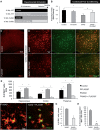
Treatment with the CSF1R inhibitor PLX3397 eliminates microglia in Rosa26-YFP mice. Two-month-old mice were treated with PLX3397 (600 mg/kg) for 7 days to eliminate microglia. (A) Schematic of the breeding strategy to yield offspring with YFP expressing microglia. (B) Immunolabelling for microglia (IBA1 in red) and expression of YFP in CSF1R+/derived cells (YFP in green). (C) Quantification of the number of YFP+ and IBA1+ cells is reduced in the cortex by 88% (P < 0.0001) and 99% (P < 0.0001), respectively, with PLX3397 treatment. Statistical significance is denoted by *P<0.05. Error bars indicate SEM (n = 4/group).
Figure 2
Figure 2
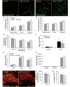
Chronically activated microglia in 5xfAD mice are dependent on CSF1R signalling for their survival. Ten-month-old wild-type or 5xfAD mice were treated with control chow or PLX3397 for 28 days to eliminate microglia. (A) Experimental design. (B) In contextual fear conditioning, 5xfAD mice spent significantly less time freezing compared to control (via two-way ANOVA, P = 0.0196) and 5xfAD + PLX3397 treated mice trended to an increased freezing time (via two-way ANOVA, P = 0.0813). (C and D) Immunolabelling for microglia (IBA1 in red) and staining for dense-core plaques (Thio-S in green). (E) Microglia number is increased by ∼40% in 5xfAD mice compared to control (via two-way ANOVA, P < 0.0001). PLX3397 treatment eliminates ∼80% of microglia in both wild-type and 5xfAD mice (via two-way ANOVA, P < 0.001). (F) Representative 63X IBA1 Thio-S immunofluorescent staining of the cortex. (G) Quantification of plaque-associated microglia reveals the number of these cells was reduced by ∼50% with PLX3397 treatment (two-tailed unpaired t-test, P < 0.0001). (H) The number of non-plaque associated microglia is reduced by ∼90% with PLX3397 treatment in 5xfAD mice (two-tailed unpaired t-test, P < 0.0001). Statistical significance is denoted by *P < 0.05 and statistical trends by #P < 0.10. Error bars indicate SEM (n = 7/group).
Figure 3
Figure 3
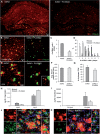
Elimination of microglia does not modulate amyloid-β levels or plaque load. (A and B) Representative hippocampal and thalamic 10× images of dense core plaques (Thio-S) in 5xfAD and 5xfAD mice treated with PLX3397. (C and D) Quantification of number of Thio-S+ plaques and average area of the plaques in the hippocampus, cortex, and thalamus. (E) Microglial elimination has no effect on plaques of any size. (F) Levels of amyloid-β1–40 and amyloid-β1–42 were unchanged with microglial elimination in wild-type mice. Levels of amyloid-β1–38 were below detection threshold. (G). (H) Levels of amyloid-β species in detergent-soluble and formic acid-soluble (FA) fractions are not changed with microglial elimination. (I) Representative 10× 6E10 and IBA1 immunofluorescent images of the thalamus. (J and K) Quantification of 6E10+ plaque size and numbers reveals no effect of microglial elimination. Error bars indicate SEM (n = 7/group).
Figure 4
Figure 4
Microglial elimination in young 5xfAD mice does not affect amyloid-β pathology. One and a half-month-old 5xfAD mice were treated with PLX3397 or control chow for 28 days. (A) Experimental design. (B and C) Representative 10× immunofluorescent IBA1 and 6E10 images of the hippocampus and amygdala. (D) Quantification of microglial cells shows a significant reduction in the 5xfAD + PLX3397 group compared to the 5xfAD group in the hippocampus and amygdala (two-tailed unpaired t-test, P < 0.0001). (E and F) Levels of amyloid-β species in detergent-soluble fractions are unchanged with the elimination of microglia. Levels of amyloid-β1–38 in the detergent-soluble fraction were below detection threshold. Statistical significance is denoted by *P < 0.05. Error bars indicate SEM (n = 4/group). FOV = field of view.
Figure 5
Figure 5
Elimination of microglia with PLX5622. Fourteen-month-old 5xfAD mice were treated with PLX5622 or control chow for 28 days. (A, B and E) Representative images showing dense-core plaques (Thio-S in green) and microglia (IBA1 in red) in the hippocampus and cortex. (C and D) Quantification reveals ∼65% decrease in plaque-associated microglia with PLX5622 treatment (two-tailed unpaired t-test; P < 0.0001) and a significant increase in specifically 0 to 1 IBA1+ cells associated with plaques in the PLX5622 group (two-tailed unpaired t-test; P < 0.0001). (F and G) Quantification of Thio-S+ plaque areas and number of 6E10+ plaques shows no effect of microglial elimination. (H and I) Levels of soluble and insoluble amyloid-β species in 5xfAD mice show no significant changes with microglial elimination. (J and K) 63X image of microglia (IBA1 in green), plaques (6E10 in red), and cell nuclei (DAPI in blue). Statistical significance is denoted by *P < 0.05. Error bars indicate SEM (n = 4/group). FOV = field of view.
Figure 6
Figure 6
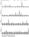
Modest changes in Alzheimer's disease-related genes with microglial elimination. Transcript levels for Alzheimer's disease-related genes were analysed using NanoString nCounter platform and immunoblots were performed to assess components of APP processing. (A) Alzheimer's disease-related gene transcript levels in all four experimental groups. Symbols denote significant differences between groups (P < 0.05): *Control versus PLX3397; Control versus 5xfAD; #PLX3397 versus 5xfAD + PLX3397; ϕ5xfAD versus 5xfAD + PLX3397. (B and C) Immunoblotting for full-length APP, C99 and C83 showed significantly increased levels of each protein in 5xfAD mice compared to Control (via two-way ANOVA; P < 0.0001) and in 5xfAD + PLX3397 mice compared to PLX3397 (via two-way ANOVA; P < 0.0001), but showed no effect of microglial elimination in 5xfAD mice. Statistical significance is denoted by *P < 0.05. Error bars indicate SEM (n = 3–6/group).
Figure 7
Figure 7
Microglial elimination reduces neuroinflammatory signalling. RNA transcripts for inflammation-related genes were analysed using NanoString nCounter platform. Symbols denote significant differences between groups (P < 0.05): symbols denote significant differences between groups (P < 0.05): *Control versus PLX3397; Control versus 5xfAD; #PLX3397 versus 5xfAD + PLX3397; ϕ5xfAD versus 5xfAD + PLX3397. Error bars indicate SEM (n = 3–6/group).
Figure 8
Figure 8
Microglia modulate dendritic spine number in the CA1 and prevent neuronal loss in the subiculum of 5xfAD mice. (A) Representative ×2.5 image of Golgi staining in hippocampus and cortex. (B) Representative ×20 image of CA1 neurons and dendritic branches. (C) Representative ×100 images of CA1 dendritic branches showing spines. (D) The number of total (two-way ANOVA; P = 0.0337) and mushroom (via two-way ANOVA; P = 0.0091) dendritic spines is significantly decreased in 5xfAD mice, compared to control. 5xfAD + PLX3397 mice show significantly increased mushroom spine density (via two-way ANOVA; P = 0.0416) and a trend for recovery of total spine loss (via two-way ANOVA; P = 0.0561), as well as a trend for increased thin spine density compared to 5xfAD (via two-way ANOVA; P = 0.066). (E) Representative ×2.5 images of Cresyl violet staining with the subiculum outlined in yellow. (F) Stereological quantification of the number of neurons in the 5xfAD group showed a significant decrease in cell number compared to control (two-way ANOVA; P = 0.0181). The neuronal loss in the 5xfAD group is prevented with microglial elimination in the 5xfAD + PLX3397 group (two-way ANOVA; P = 0.0458). Statistical significance is denoted by *P < 0.05 and statistical trends by #P < 0.10. Error bars indicate SEM (n = 3–4/group).

References

    1. Akama KT, Van Eldik LJ.Beta-amyloid stimulation of inducible nitric-oxide synthase in astrocytes is interleukin-1beta- and tumor necrosis factor-alpha (TNFalpha)-dependent, and involves a TNFalpha receptor-associated factor- and NFkappaB-inducing kinase-dependent signaling mechanism. J Biol Chem 2000; 275: 7918–24. - PubMed
    1. Akiyama H, Arai T, Kondo H, Tanno E, Haga C, Ikeda K. Cell mediators of inflammation in the Alzheimer disease brain. Alzheimer Dis Assoc Disord 2000; 14 (Suppl 1): S47–53. - PubMed
    1. Ando K, Brion JP, Stygelbout V, Suain V, Authelet M, Dedecker R, et al. Clathrin adaptor CALM/PICALM is associated with neurofibrillary tangles and is cleaved in Alzheimer's brains. Acta Neuropathol 2013; 125: 861–78. - PubMed
    1. Asai H, Ikezu S, Tsunoda S, Medalla M, Luebke J, Haydar T, et al. Depletion of microglia and inhibition of exosome synthesis halt tau propagation. Nat Neurosci 2015; 18: 1584–93. - PMC - PubMed
    1. Blinzinger K, Kreutzberg G. Displacement of synaptic terminals from regenerating motoneurons by microglial cells. Z Zellforsch Mikrosk Anat 1968; 85: 145–57. - PubMed

Publication types

Substances