Review
. 2016 Jul:109:119-31.
doi: 10.1016/j.phrs.2016.02.020. Epub 2016 Feb 26.

Acetaminophen from liver to brain: New insights into drug pharmacological action and toxicity

Affiliations
Review

Acetaminophen from liver to brain: New insights into drug pharmacological action and toxicity

Carolina I Ghanem et al. Pharmacol Res. 2016 Jul.

Abstract

Acetaminophen (APAP) is a well-known analgesic and antipyretic drug. It is considered to be safe when administered within its therapeutic range, but in cases of acute intoxication, hepatotoxicity can occur. APAP overdose is the leading cause of acute liver failure in the northern hemisphere. Historically, studies on APAP toxicity have been focused on liver, with alterations in brain function attributed to secondary effects of acute liver failure. However, in the last decade the pharmacological mechanism of APAP as a cannabinoid system modulator has been documented and some articles have reported "in situ" toxicity by APAP in brain tissue at high doses. Paradoxically, low doses of APAP have been reported to produce the opposite, neuroprotective effects. In this paper we present a comprehensive, up-to-date overview of hepatic toxicity as well as a thorough review of both toxic and beneficial effects of APAP in brain.

Keywords: Acetaminophen; Caspase; DAMPs; Hepatic toxicity; Necroptosis; trpv1.


Figures

Figure 1
Figure 1. FAAH-metabolite of APAP: Generation of N-arachidonoyl-phenolamine (AM404) in Brain
Panel A: APAP is deacetylated followed by the formation of AM404 by the addition of arachidonic acid, which is catalyzed by fatty acid amide hydrolase (FAAH) in brain. Panel B: Structures of AM404 and
Figure 2
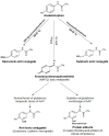
Figure 2. Liver APAP Metabolism
Main metabolic pathways of APAP in liver after administration of therapeutic or toxic doses.
Figure 3
Figure 3. Mechanism of APAP Toxicity in Liver
An Excess of the reactive intermediate NAPQI after GSH stores are depleted is considered to be the molecular initial event in APAP toxicity. Unneutralized NAPQI forms covalent adducts with macromolecules in hepatocytes, increases oxidative stress and inhibits Ca2+-Mg2+-ATPases, which results in intracellular Ca2+ dysregulation and accumulation. This increase in Ca2+ can activate calpains, mediating proteolytic degradation. Mitochondrial GSH depletion and selective mitochondrial proteins adducts are associated with ROS generation and formation of membrane permeability transition (MPT) pore, which triggers mitochondrial damage and release of cit c. Inhibition of mitochondrial respiration decreases ATP levels, which in terms halt the apoptotic signaling cascade and redirects the cell fate toward necrosis. After initial formation of NAPQI and adducts, several other molecular events contribute to the amplification and propagation of tissue damage. Recently, the phosphorylation of JNK by MAPKK and activation of RIPK1 through necroptosis or by direct interaction with JNK has been also implicated in APAP cytotoxicity. Abbreviations: APAP, acetaminophen; AIF, apoptosis inducing factor; ATP, adenosine triphosphate; CYP, cytochrome P-450 isoenzymes mixed-function oxidase system;Cyt c, cytochrome c; DNA, Deoxyribonucleic acid; Fe2+ iron, GSH, GlutathioneJNK, c-Jun N-terminal kinase; MAPK, mitogen-Activated; MPT, membrane permeability transition; NAPQI, N-acetyl-p-benzoquinoneimine; Nec, necrostatins; NO, nitric oxide; O2-, superoxide; RIP, receptor interacting protein kinase; ROS, reactive oxygen species; Sab, Src homology 3 domain binding protein 5; Δψ m, mitochondrial membrane potential.
Figure 4
Figure 4. Time-dependent Effect of APAP on Brain Nrf2 Gene Expression and its Nuclear Localization
Brain Nrf2 mRNA levels were measured by qPCR. RNA isolation and quantification was performed as detailed in Ghanem et al. (2015) (126). Data are presented as percentage of control and expressed as means ± SE (n=5 mice/group). Nrf2 protein nuclear concentration was determined by Western blotting. Nuclear protein and western-blot was performed as was described in Ghanem et al. (2015) (126). Equal protein (50 μg) was loaded into each lane. TATA-binding protein (TBP) was used as loading control. Densitometric analysis of blot is presented as percentage of control and expressed as mean ± SE (n=5 mice/group. * p<0.05 vs Oh).
Figure 5
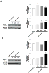
Figure 5. Time-dependent Effect of APAP on the Expression of the Nrf2 Target Genes Ho-1 and Nqo1
Brain Ho-1 (Panel A) and Nqo1 (Panel B) mRNA and protein levels were measured by qPCR and Western blotting, respectively. Total homogenates were used and the methodology used in RNA and protein studies are detailed in Ghanem et al. (2015) (126). mRNA data are presented as percentage of control and expressed as mean ± SE. For western blotting, equal cytosolic protein (50 μg) was loaded into each lane. β-actin was used as loading control. Densitometric analysis of blots is presented as percentage of control values and expressed as mean ± SE. (n=5 mice/group. *** p<0.001; ** p<0.01; * p<0.05 vs 0h).
Figure 6
Figure 6. Effect of Different Concentrations of APAP on Primary Culture of Glial Cells
Glial primary cultures from newborn Wistar rats were performed according to Pérez el al (2013) (138). Cell cultures were astrocytes (80–85%) and oligodendrocyte (15–20%). Panel A: Cell viability. The cell viability of glial primary culture was measured by the MTT assay (139) after incubation with APAP (1, 5, 10, 20 mM) for 48 h. The absorbance at 570nm was expressed as percentage of control. Panel B: Cell proliferation. Proliferation was evaluated in glial primary cultures using 1 and 20mM APAP by Whole-cell BdrU ELISA as was described by Silvestroff et al. (2012) (139). The results were expressed as absorbance at 450nm. Panel C: Cell death. Propidium iodine (PI) staining was used to identify dead cells. Total cells were stained with hoechst (139). Data in A, B and C are means±SD of four independent cultures. One-way ANOVA followed by Newman-Keuls multiple comparison test were used in A, B, and C to determine statistical significance; ***P< 0.001, *P< 0.05, symbols above the bar indicate significance compared to corresponding control.

References

    1. Morse HN. Ueber eine neue Darstellungsmethode der Acetylamidophenole. Ber Deutscher Chem Ges. 1878;11:232–233.
    1. Von Mering J. Beitrage zur Kenntniss der Antipyretica. Ther Monatsch. 1893;7:577–587.
    1. Brodie BB, AXELROD J. The fate of acetanilide in man. J Pharmacol Exp Ther. 1948;94:29–38. - PubMed
    1. Bentley E, Mackie IC. Trends in prescriptions of paracetamol for children. BMJ. 1995;311:362. - PMC - PubMed
    1. Pandolfini C, Bonati M. A literature review on off-label drug use in children. Eur J Pediatr. 2005;164:552–558. - PubMed

Publication types

MeSH terms