Review
. 2016 Apr;18(4):25.
doi: 10.1007/s11912-016-0506-0.

What Is the Best Preoperative Imaging for Endometrial Cancer?

Affiliations
Review

What Is the Best Preoperative Imaging for Endometrial Cancer?

Ingfrid S Haldorsen et al. Curr Oncol Rep. 2016 Apr.

Abstract

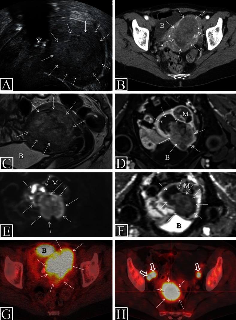
Although endometrial cancer is surgicopathologically staged, preoperative imaging is recommended for diagnostic work-up to tailor surgery and adjuvant treatment. For preoperative staging, imaging by transvaginal ultrasound (TVU) and/or magnetic resonance imaging (MRI) is valuable to assess local tumor extent, and positron emission tomography-CT (PET-CT) and/or computed tomography (CT) to assess lymph node metastases and distant spread. Preoperative imaging may identify deep myometrial invasion, cervical stromal involvement, pelvic and/or paraaortic lymph node metastases, and distant spread, however, with reported limitations in accuracies and reproducibility. Novel structural and functional imaging techniques offer visualization of microstructural and functional tumor characteristics, reportedly linked to clinical phenotype, thus with a potential for improving risk stratification. In this review, we summarize the reported staging performances of conventional and novel preoperative imaging methods and provide an overview of promising novel imaging methods relevant for endometrial cancer care.

Keywords: Computed tomography; Diffusion weighted imaging; Endometrial cancer; Imaging biomarkers; Magnetic resonance imaging; Positron emission tomography; Preoperative imaging; Staging; Vaginal ultrasound.


Figures

Fig. 1
Fig. 1
Characteristic preoperative imaging findings in endometrial cancer. VUS (a) in patient with FIGO stage 1B (endometrioid, grade 2) depicting a large uterine tumor (arrows) with mixed echogenicity and signs of deep myometrial invasion. CE CT (b), sagittal T2 weighted MRI (c), axial CE T1-weighted MRI (d), axial DWI (b = 1000 s/mm2) (e) with ADC map (f), and FDG PET-CT (g) in a patient with FIGO stage 1B (endometrioid, grade 3). The large uterine tumor (arrows), invading >50 % of the myometrial wall, is hypodense relative to the surrounding myometrium at CT (b), hyperintense at T2-weighted MRI (c), and hypointense at CE T1-weighted MRI. DWI shows tumor hyperintensity (e) with corresponding hypointensity on the ADC map (f; mean ADC value of 0.54 × 10−3 mm2/s), indicating restricted diffusion within the tumor. The same lesion is FDG avid at FDG PET-CT (g; SUVmax of 10.4). FDG PET-CT in patient with FIGO stage 3C2 (endometrioid, grade 3) (h) depicts large FDG avid tumor (SUVmax of 25.0) and three pelvic lymph node metastases (h, open arrows; SUVmax of 11.3). The bladder (b) normally appears FDG avid due to FDG secretion in the urine (g). Note the concomitant calcified myoma (m) seen at VUS (a) and the adjoining myoma (m) depicted at MRI (df) with no restricted diffusion; the myoma is thus easy to differentiate from the uterine tumor. ADC apparent diffusion coefficient, B bladder, CE contrast enhanced, CT computed tomography, DWI diffusion weighted imaging, FDG fluorodeoxyglucose, M myoma, MRI magnetic resonance imaging, PET positron emission tomography, SUV standard uptake value, VUS vaginal ultrasound

References

    1. Amant F, Moerman P, Neven P, Timmerman D, Van LE, Vergote I. Endometrial cancer. Lancet. 2005;366(9484):491–505. doi: 10.1016/S0140-6736(05)67063-8. - DOI - PubMed
    1. Salvesen HB, Haldorsen IS, Trovik J. Markers for individualised therapy in endometrial carcinoma. Lancet Oncol. 2012;13(8):e353–61. doi: 10.1016/S1470-2045(12)70213-9. - DOI - PubMed
    1. Creasman WT, Odicino F, Maisonneuve P, Quinn MA, Beller U, Benedet JL, et al. Carcinoma of the corpus uteri. FIGO 6th annual report on the results of treatment in gynecological cancer. Int J Gynaecol Obstet. 2006;95(Suppl 1):S105–43. doi: 10.1016/S0020-7292(06)60031-3. - DOI - PubMed
    1. Oza AM, Elit L, Tsao MS, Kamel-Reid S, Biagi J, Provencher DM, et al. Phase II study of temsirolimus in women with recurrent or metastatic endometrial cancer: a trial of the NCIC Clinical Trials Group. J Clin Oncol. 2011;29(24):3278–85. doi: 10.1200/JCO.2010.34.1578. - DOI - PMC - PubMed
    1. Pecorelli S. Revised FIGO staging for carcinoma of the vulva, cervix, and endometrium. Int J Gynaecol Obstet. 2009;105(2):103–4. doi: 10.1016/j.ijgo.2009.02.012. - DOI - PubMed

Publication types

MeSH terms

LinkOut - more resources