. 2016 Dec;6(1):8.
doi: 10.1186/s12348-016-0074-3. Epub 2016 Feb 27.

Vanishing retinal arterial aneurysms with anti-tubercular treatment in a patient presenting with idiopathic retinal vasculitis, aneurysms, and neuroretinitis

Affiliations

Vanishing retinal arterial aneurysms with anti-tubercular treatment in a patient presenting with idiopathic retinal vasculitis, aneurysms, and neuroretinitis

Ramandeep Singh et al. J Ophthalmic Inflamm Infect. 2016 Dec.

Abstract

Background: Idiopathic retinal vasculitis, aneurysms, and neuroretinitis (IRVAN) syndrome presents with characteristic clinical manifestations such as aneurysms at arteriolar bifurcations and optic nerve and retinal vascular inflammation. Regression of such features on treatment with anti-tubercular therapy (ATT) combined with corticosteroids has not been reported in literature.

Findings: A 30-year-old female with sudden painless decreased vision in the left eye was referred with a diagnosis of presumed tuberculous retinal vasculitis and a positive tuberculin skin test. Based on the clinical and angiographic features of the right eye, a diagnosis of IRVAN syndrome was made. In the left eye, the patient had vitreous hemorrhage for which pars plana vitrectomy was performed. The vitreous sample was positive for Mycobacterium tuberculosis using multiplex polymerase chain reaction, and the patient was started on standard four-drug ATT and oral corticosteroids. At 6-month follow-up, vanishing of retinal arterial aneurysms was observed.

Conclusions: The pathogenesis of IRVAN syndrome is uncertain. One of the postulates is that the features of arterial aneurysms and other retinal vascular alterations occur secondary to acquired inflammatory reaction. We hypothesize that IRVAN syndrome may be a morphological diagnosis possibly associated with various entities, one of which could be ocular tuberculosis. It may be prudent to rule out intraocular tuberculosis in cases labeled as IRVAN syndrome in an endemic population.

Keywords: Aneurysms; Anti-tubercular therapy; Fluorescein angiography; IRVAN; Intraocular tuberculosis; Polymerase chain reaction; Uveitis.


Figures

Fig. 1
Fig. 1
Fundus photograph (a) and fluorescein angiogram (FA) (bd) of the right eye at presentation showing hard exudates, superficial hemorrhages, and “tied-knot-like” aneurysmal dilatations of the arterioles on optic disc and in the surrounding region. In addition, vascular sheathing can be observed in the larger vessels temporally. A large area of capillary non-perfusion can be seen temporally on FA (b). In the late phase (d), significant leakage can be observed from the peripheral vessels at the junction of perfused and non-perfused retina
Fig. 2
Fig. 2
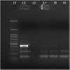
Positive results of multiplex polymerase chain reaction demonstrating positivity to tubercular antigens in the vitreous fluid. Band L1 represents 100-bp molecular marker; L2 is the positive control (asterisk indicates protein B (419 bp), double asterisks indicate MPB64 (240 bp), dagger indicates IS6110 (123 bp)). Band L3 is the negative control. Bands L4, L5, and L6 represent the positive results obtained from the vitreous fluid of the patient described in the index case
Fig. 3
Fig. 3
Comparison of the posterior pole fundus photographs at baseline (a), 6 months (b), and 11 months (c) of the right eye posterior pole (magnified) shows resolution of arterial aneurysms with resolution of hard exudates. Fluorescein angiogram (FA) at baseline (d) versus 6 month (e) and 11 month (f) shows much reduced aneurysmal dilations at arteriolar branching (yellow arrows). Laser scars of pan-retinal photocoagulation are clearly demonstrated in the FA at month 6 superiorly (e)

References

    1. Chang TS, Aylward GW, Davis JL, Mieler WF, Oliver GR, Maberley AL, Gass JD. Idiopathic retinal vasculitis, aneurysms, and neuroretinitis: retinal vasculitis study. Ophthalmology. 1995;102:1089–97. doi: 10.1016/S0161-6420(95)30907-4. - DOI - PubMed
    1. Kincaid J, Schatz H. Bilateral retinal arteritis with multiple aneurysmal dilatations. Retina. 1983;3:171–8. doi: 10.1097/00006982-198300330-00005. - DOI - PubMed
    1. Karagiannis D, Soumplis V, Georgalas I, Kandarakis A. Ranibizumab for idiopathic retinal vasculitis, aneurysms, and neuroretinitis: favorable results. Eur J Ophthalmol. 2010;20:792–4. - PubMed
    1. Cheema RA, Al-Askar E, Cheema HR. Infliximab therapy for idiopathic retinal vasculitis, aneurysm, and neuroretinitis syndrome. J Ocul Pharmacol Ther. 2011;27:407–10. doi: 10.1089/jop.2010.0189. - DOI - PubMed
    1. Abu El-Asrar AM, Jestaneiah S, Al-Serhani AM. Regression of aneurysmal dilatations in a case of idiopathic retinal vasculitis, aneurysms and neuroretinitis (IRVAN) associated with allergic fungal sinusitis. Eye. 2004;18:197–201. doi: 10.1038/sj.eye.6700554. - DOI - PubMed

LinkOut - more resources