. 2016 Feb 29:6:22263.
doi: 10.1038/srep22263.

Isothiocyanates as effective agents against enterohemorrhagic Escherichia coli: insight to the mode of action

Affiliations

Isothiocyanates as effective agents against enterohemorrhagic Escherichia coli: insight to the mode of action

Dariusz Nowicki et al. Sci Rep. .

Abstract

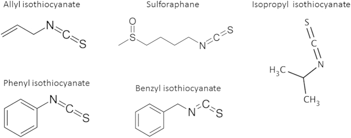
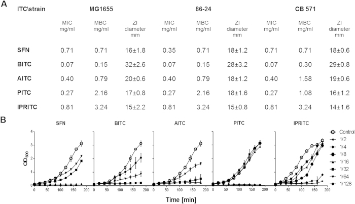
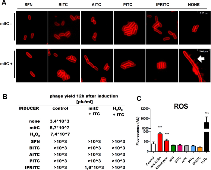
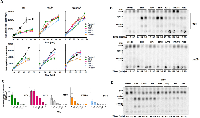
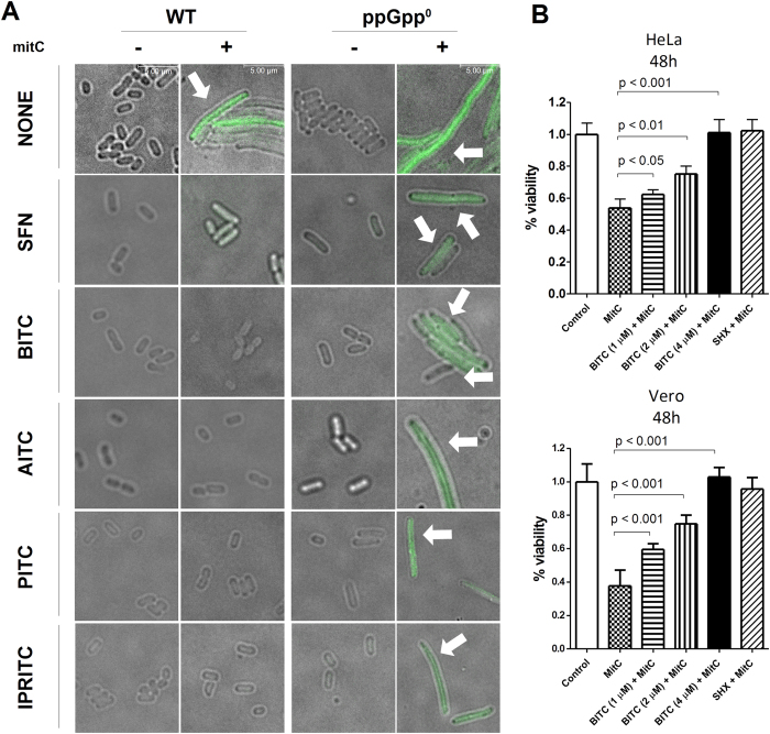
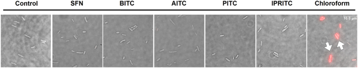
Production of Shiga toxins by enterohemorrhagic Escherichia coli (EHEC) which is responsible for the pathogenicity of these strains, is strictly correlated with induction of lambdoid bacteriophages present in the host's genome, replication of phage DNA and expression of stx genes. Antibiotic treatment of EHEC infection may lead to induction of prophage into a lytic development, thus increasing the risk of severe complications. This, together with the spread of multi-drug resistance, increases the need for novel antimicrobial agents. We report here that isothiocyanates (ITC), plant secondary metabolites, such as sulforaphane (SFN), allyl isothiocyanate (AITC), benzyl isothiocynanate (BITC), phenyl isothiocyanate (PITC) and isopropyl isothiocyanate (IPRITC), inhibit bacterial growth and lytic development of stx-harboring prophages. The mechanism underlying the antimicrobial effect of ITCs involves the induction of global bacterial stress regulatory system, the stringent response. Its alarmone, guanosine penta/tetraphosphate ((p)ppGpp) affects major cellular processes, including nucleic acids synthesis, which leads to the efficient inhibition of both, prophage induction and toxin synthesis, abolishing in this way EHEC virulence for human and simian cells. Thus, ITCs could be considered as potential therapeutic agents in EHEC infections.


Figures

Figure 1
Figure 1. The chemical structures of ITCs employed in this study.
Figure 2
Figure 2. Effect of ITCs on bacterial growth.
(A) MIC, MBC and zone inhibition were determined for each ITC. The results are from at least three independent experiments (B) E. coli strain MG1655 was cultivated in the absence or in the presence of ITCs (SFN, BITC, AITC, PITC, IPRITC). Concentrations of ITC are presented in relation to MIC determined as in A.
Figure 3
Figure 3. ITCs affect the prophage induction.
The effects of ITCs and prophage induction by mitomycin C or hydrogen peroxide were assessed by the microscopic analysis (A), by phage yield (B) and by production of reactive oxygen species (C). (A) For microscopic analysis, prophage induction was provoked by addition of mitomycin C (1 μg/ml), ITCs (as indicated) were added at the same time. After 3 hours, samples were stained with SynaptoRed to visualize bacterial membranes. The white arrows mark filamentous E. coli cells, (B) phage yield was measured 12 hours after induction by mitomycin C (1 μg/ml) or hydrogen peroxide (1 μM). ITCs at the concentrations of 1/8, as determined for each ITC, were added at the time of induction, (C) reactive oxygen species level was assessed after prophage induction with hydrogen peroxide, or presence of indicated ITCs or antibiotics. The experiments were repeated independently at least 3 times. The differences among the results were examined by the T-student test. Differences with statistical significance are indicated by asterisk above columns indicating P value < 0.001.
Figure 4
Figure 4. Effect of ITCs on nucleic acids metabolism.
(A) DNA and RNA synthesis were assessed in E. coli wild type, ΔrelA and ΔrelA ΔspoT (ppGpp0) strains (as indicated above each panel) in the absence (empty circles) or presence of ITCs: SFN (green), BITC (magenta), AITC (grey), IPRITC (yellow), PITC (light blue) at subinhibitory concentrations (1/32,1/16,1/16,1/8,1/8 MIC respectively). The relative nucleic acid synthesis was measured by the level of incorporation of radioactive-labeled substrates, [3H]thymidine or [3H]uridine and presented as counts per minute (cpm) normalized to cell growth (A600). ITCs were added at time zero. The results are mean values from three independent experiments done in duplicate, with error bars indicating SD. (B) The synthesis of the stringent response alarmones, ppGpp and pppGpp was assessed by culturing the the wild type or relA bacteria in the presence of [32P]orthophosphoric acid (150 μCi/ml) followed by cell lysis and nucleotide separation by thin-layer chromatography. ITCs or SHX were added at time zero. Samples were withdrawn at 15 and 30 min after the addition. The positions of ppGpp and pppGpp are indicated by arrows. (C) The relative (p)ppGpp synthesis at 30 min after addition of indicated ITCs quantified by densitometry using phosphorimager (Typhoon, GE Healthcare) and Quantity One software. The results are the average of three independent experiments ± SD, normalized by setting the ppGpp level obtained in SHX treatment as 1. (D) The (p)ppGpp synthesis was assessed as in (B) with the addition of the amino acids (final concentration at 400 μg/ml) where indicated.
Figure 5
Figure 5. ITCs inhibit stx gene expression and toxicity of EHEC lysates.
(A) The prophage induction by mitomycin C (1 μg/ml) in the presence or absence of ITCs (as indicated) was assessed for E. coli wild type and ppGpp0 strains lysogenized with 933 W phage. GFP fluorescence corresponds to the expression of stx genes (indicated by arrow). (B) Effect of BITC and SHX on viability of HeLa and Vero cells treated with EHEC strain lysate. The Shiga-toxin harboring prophage was induced in E. coli 86–24 strain by mitomycin C (1 μg/ml) in the absence (control) or presence of various BITC concentrations or 0.5 mg/ml SHX.
Figure 6
Figure 6. The effect of ITCs on cellular membrane integrity.
Bacteria were treated with indicated ITCs at growth inhibitory concentrations, and with propidium iodide; the fluorescence signal was visualized by fluorescence microscopy. Cells with damaged membranes were marked with white arrows.

Similar articles

Cited by

References

    1. Howie H., Mukerjee A., Cowden J., Leith J. & Reid T. Investigation of an outbreak of Escherichia coli O157 infection caused by environmental exposure at a scout camp. Epidemiol. Infect. 131, 1063–9 (2003). - PMC - PubMed
    1. Beutin L. & Martin A. Outbreak of Shiga toxin-producing Escherichia coli (STEC) O104:H4 infection in Germany causes a paradigm shift with regard to human pathogenicity of STEC strains. J. Food Prot. 75, 408–418 (2012). - PubMed
    1. Bloch S. A., Felczykowska A. & Nejman-Faleńczyk B. Escherichia coli O104:H4 outbreak-have we learnt a lesson from it? Acta Biochim. Pol. 59, 483–488 (2012). - PubMed
    1. Werber D. et al. Outbreaks of virulent diarrheagenic Escherichia coli—are we in control? BMC Med. 10, 11 (2012). - PMC - PubMed
    1. Yahata Y. et al. Epidemiological analysis of a large enterohaemorrhagic Escherichia coli O111 outbreak in Japan associated with haemolytic uraemic syndrome and acute encephalopathy. Epidemiol. Infect. 20, 1–12 (2015). - PMC - PubMed

Publication types

MeSH terms