Review
. 2016 Oct 13;35(41):5337-5349.
doi: 10.1038/onc.2016.34. Epub 2016 Feb 29.

The tumor microenvironment in esophageal cancer

Affiliations
Review

The tumor microenvironment in esophageal cancer

E W Lin et al. Oncogene. .

Abstract

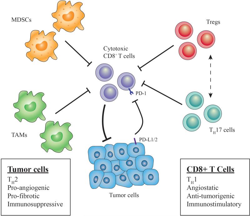
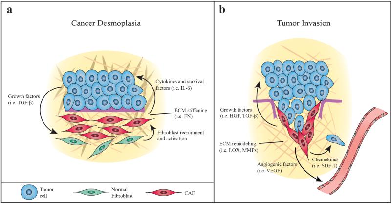
Esophageal cancer is a deadly disease, ranking sixth among all cancers in mortality. Despite incremental advances in diagnostics and therapeutics, esophageal cancer still carries a poor prognosis, and thus, there remains a need to elucidate the molecular mechanisms underlying this disease. There is accumulating evidence that a comprehensive understanding of the molecular composition of esophageal cancer requires attention to not only tumor cells but also the tumor microenvironment (TME), which contains diverse cell populations, signaling factors and structural molecules that interact with tumor cells and support all stages of tumorigenesis. In esophageal cancer, environmental exposures can trigger chronic inflammation, which leads to constitutive activation of pro-inflammatory signaling pathways that promote survival and proliferation. Antitumor immunity is attenuated by cell populations such as myeloid-derived suppressor cells and regulatory T cells, as well as immune checkpoints like programmed death-1. Other immune cells such as tumor-associated macrophages can have other pro-tumorigenic functions, including the induction of angiogenesis and tumor cell invasion. Cancer-associated fibroblasts secrete growth factors and alter the extracellular matrix to create a tumor niche and enhance tumor cell migration and metastasis. Further study of how these TME components relate to the different stages of tumor progression in each esophageal cancer subtype will lead to development of novel and specific TME-targeting therapeutic strategies, which offer considerable potential especially in the setting of combination therapy.


Figures

Figure 1
Figure 1. Immune landscape in esophageal cancer
Several immune cell types disrupt anti-tumor immunity (cytotoxic CD8+ T cells) in the tumor microenvironment (TME). Tregs expressing CCR4 are recruited by chemokines CCL17 and CCL22 that are secreted by tumor cells (and TAMs). Tregs exert immunosuppressive function via direct contact with effector T cells or by molecules such as adenosine or immunosuppressive cytokines (IL-10, IL-35). Th17 cells are stimulated by TGF-β and IL-6 and have the ability to convert into Tregs (dashed line) and release adenosine by ectoenzymatic (CD39, CD73) function. Expansion of myeloid derived suppressor cells (MDSC), or immature myeloid cells, is stimulated by inflammation and tumor-derived factors (i.e. VEGF), and these cells directly inhibit T cell activation and NK cell cytotoxicity, while also inducing Tregs. TAM expansion (M2 polarization) occurs in presence of Th2 cytokines (i.e. IL-4, IL-13), and these cells are recruited via chemokines such as MCP-1. Furthermore, TAMs and tumor cells both express PD-L1/2 to inhibit T cell activation via the PD-1 receptor. Altogether, these cells suppress anti-tumor immunity while also promoting tumor growth and progression by various mechanisms.
Figure 2
Figure 2. Stromal compartment of the esophageal TME
A. Neoplastic cells secrete growth factors to activate quiescient fibroblasts designated as cancer associated fibroblasts (CAFs). CAFs can proliferate to contribute to desmoplasia, secreting extracellular matrix (ECM) components such as fibronectin (FN) to enhance the development of the primary tumor niche. CAFs also secrete cytokines that promote tumor cell survival (anti-apoptosis). B. Later in tumorigenesis, CAFs remodel the ECM with enzymes like lysyl oxidase (LOX) and matrix metalloproteinases (MMPs) as well as ECM components like dermatan sulfate (DS) and hyaluronan (HA) to promote invasion. CAFs also secrete growth factors that trigger tumor cells to undergo epithelial-mesenchymal transition (EMT) and chemokines that induce tumor cell migration. CAFs can also promote angiogenesis via VEGF secretion.

References

    1. Pennathur A, Gibson MK, Jobe BA, Luketich JD. Oesophageal carcinoma. Lancet. 2013;381:400–12. - PubMed
    1. Rustgi AK, El-Serag HB. Esophageal Carcinoma. N Engl J Med. 2014;371:2499–2509. - PubMed
    1. Sunpaweravong P, Sunpaweravong S, Puttawibul P, Mitarnun W, Zeng C, Barón AE, et al. Epidermal growth factor receptor and cyclin D1 are independently amplified and overexpressed in esophageal squamous cell carcinoma. J Cancer Res Clin Oncol. 2005;131:111–9. - PMC - PubMed
    1. Hollstein MC, Metcalf RA, Welsh JA, Montesano R, Harris CC. Frequent mutation of the p53 gene in human esophageal cancer. Proc Natl Acad Sci U S A. 1990;87:9958–61. - PMC - PubMed
    1. Chung Y, Lam AKY, Luk JM, Law S, Chan K-W, Lee P-Y, et al. Altered E-Cadherin Expression and p120 Catenin Localization in Esophageal Squamous Cell Carcinoma. Ann Surg Oncol. 2007;14:3260–3267. - PubMed

MeSH terms