

Multi-nucleate retinal pigment epithelium cells of the human macula exhibit a characteristic and highly specific distribution
- PMID: 26923500
- PMCID: PMC4798426
- DOI: 10.1017/S0952523815000310
Multi-nucleate retinal pigment epithelium cells of the human macula exhibit a characteristic and highly specific distribution
Abstract
Background: The human retinal pigment epithelium (RPE) is reportedly 3% bi-nucleated. The importance to human vision of multi-nucleated (MN)-RPE cells could be clarified with more data about their distribution in central retina.
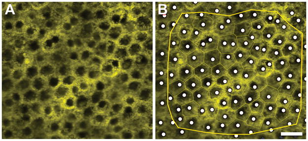
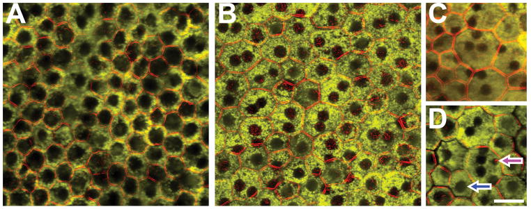
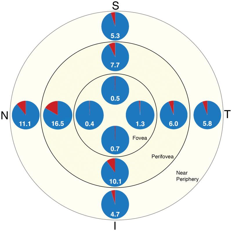
Methods: Nineteen human RPE-flatmounts (9 ≤ 51 years, 10 > 80 years) were imaged at 12 locations: 3 eccentricities (fovea, perifovea, near periphery) in 4 quadrants (superior, inferior, temporal, nasal). Image stacks of lipofuscin-attributable autofluorescence and phalloidin labeled F-actin cytoskeleton were obtained using a confocal fluorescence microscope. Nuclei were devoid of autofluorescence and were marked using morphometric software. Cell areas were approximated by Voronoi regions. Mean number of nuclei per cell among eccentricity/quadrant groups and by age were compared using Poisson and binominal regression models.
Results: A total of 11,403 RPE cells at 200 locations were analyzed: 94.66% mono-, 5.31% bi-, 0.02% tri-nucleate, and 0.01% with 5 nuclei. Age had no effect on number of nuclei. There were significant regional differences: highest frequencies of MN-cells were found at the perifovea (9.9%) and near periphery (6.8%). The fovea lacked MN-cells almost entirely. The nasal quadrant had significantly more MN-cells compared to other quadrants, at all eccentricities.
Conclusion: This study demonstrates MN-RPE cells in human macula. MN-cells may arise due to endoreplication, cell fusion, or incomplete cell division. The topography of MN-RPE cells follows the topography of photoreceptors; with near-absence at the fovea (cones only) and high frequency at perifovea (highest rod density). This distribution might reflect specific requirements of retinal metabolism or other mechanisms addressable in further studies.
Keywords: Bi-nucleate; Cell nucleus; Multi-nucleate; Retinal pigment epithelium.
Conflict of interest statement
None of the authors has any potential conflict to report.
Figures
References
-
- Ablonczy Z, Higbee D, Anderson DM, Dahrouj M, Grey AC, Gutierrez D, Koutalos Y, Schey KL, Hanneken A, Crouch RK. Lack of Correlation Between the Spatial Distribution of A2E and Lipofuscin Fluorescence in the Human Retinal Pigment Epithelium. Investigative ophthalmology & visual science. 2013;54:5535–5542. - PMC - PubMed
-
- Al-Hussaini H, Schneiders M, Lundh P, Jeffery G. Drusen are associated with local and distant disruptions to human retinal pigment epithelium cells. Experimental eye research. 2009;88:610–612. - PubMed
Publication types
MeSH terms
Grants and funding
LinkOut - more resources
Full Text Sources
Other Literature Sources
