. 2016 Mar 8;14(9):2154-2165.
doi: 10.1016/j.celrep.2016.02.004. Epub 2016 Feb 25.

Fatty Acid Oxidation-Driven Src Links Mitochondrial Energy Reprogramming and Oncogenic Properties in Triple-Negative Breast Cancer

Affiliations

Fatty Acid Oxidation-Driven Src Links Mitochondrial Energy Reprogramming and Oncogenic Properties in Triple-Negative Breast Cancer

Jun Hyoung Park et al. Cell Rep. .

Abstract

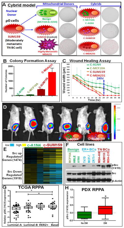
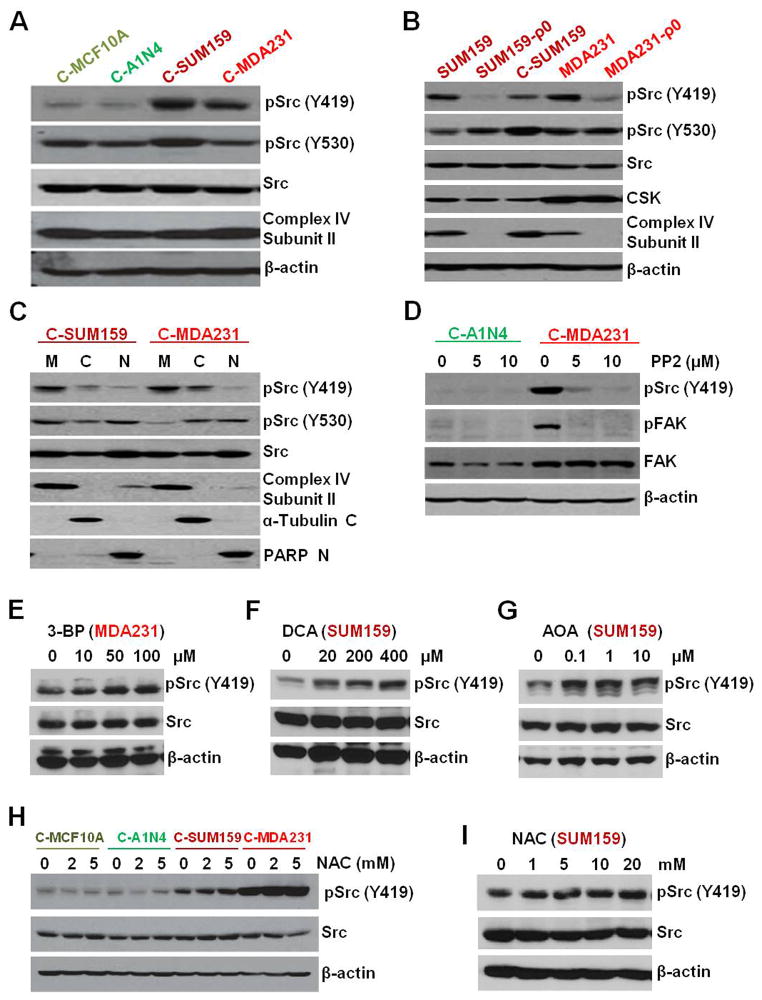
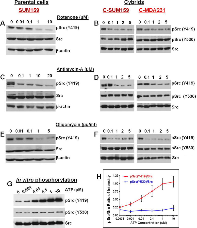
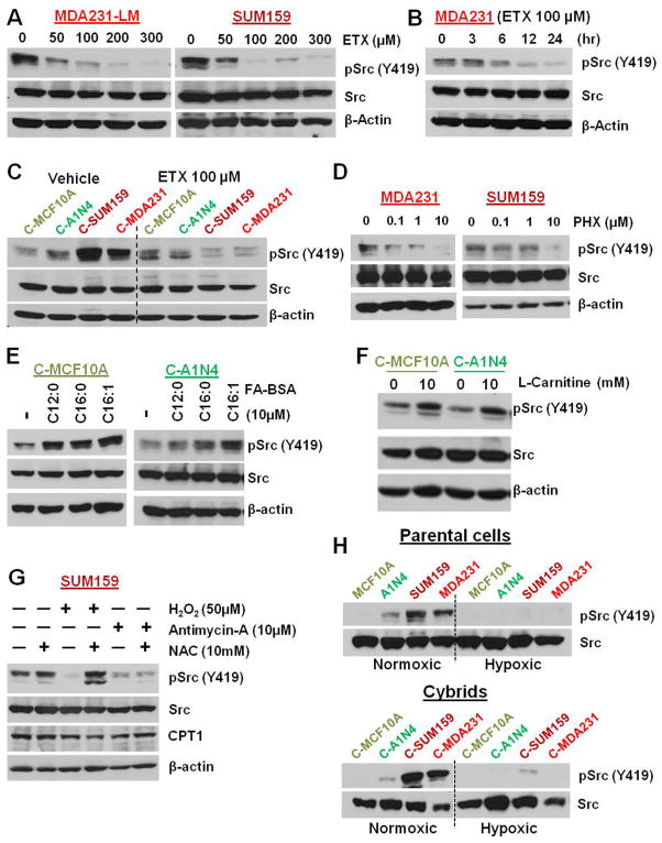
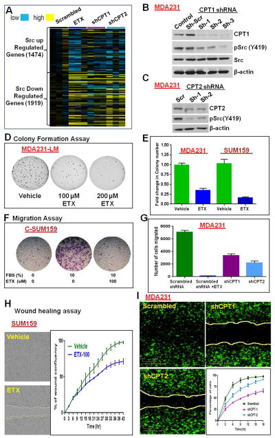
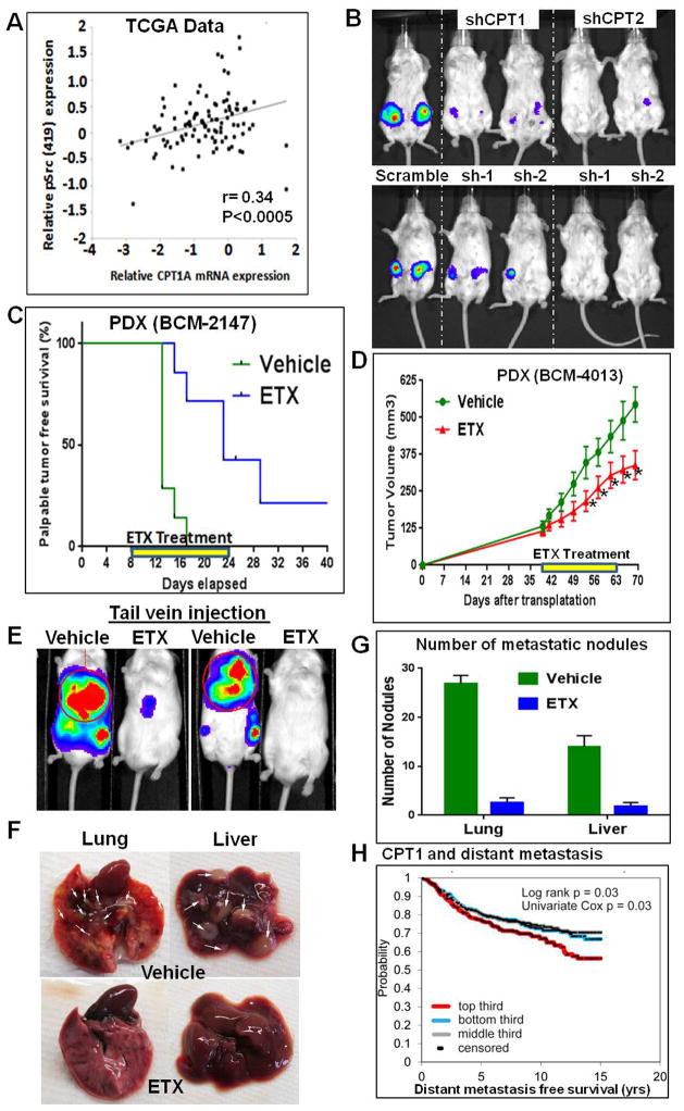
Transmitochondrial cybrids and multiple OMICs approaches were used to understand mitochondrial reprogramming and mitochondria-regulated cancer pathways in triple-negative breast cancer (TNBC). Analysis of cybrids and established breast cancer (BC) cell lines showed that metastatic TNBC maintains high levels of ATP through fatty acid β oxidation (FAO) and activates Src oncoprotein through autophosphorylation at Y419. Manipulation of FAO including the knocking down of carnitine palmitoyltransferase-1A (CPT1) and 2 (CPT2), the rate-limiting proteins of FAO, and analysis of patient-derived xenograft models confirmed the role of mitochondrial FAO in Src activation and metastasis. Analysis of TCGA and other independent BC clinical data further reaffirmed the role of mitochondrial FAO and CPT genes in Src regulation and their significance in BC metastasis.


Figures

Figure 1
Figure 1
(A) SUM159 TNBC cybrid model with mitochondria from benign (MCF10A and A1N4), moderately metastatic (SUM159), and highly metastatic (MDA231) TN cells under a defined nuclear background of SUM159 mitochondrial DNA depleted ρ0 cells. The cybrids are named as C-MCF10A, C-A1N4, C-SUM159, and C-MDA231 respectively. Images of soft agar colony formation assay of mitochondrial donor cells and cybrids are shown with the cartoon. Colonies are red marked using GelCount software. (B) Quantification of soft agar colonies of cybrids. (C) Analysis of wound healing assays performed in an IncuCyte ZOOM® kinetic imaging system with live-cell imaging every three hours. While cybrids with mitochondria from cancer cells completely healed (100% relative wound density) within 24 hours, cybrids with mitochondria from benign cells could not heal the wound even after 36 hours. (D) Bioluminescence imaging of tumor growth in cybrids. Mice injected in the mammary fat pads with cybrids C-A1N4 [right (R); cartooned with green mitochondria] and C-SUM159 [left (L); cartooned with pink mitochondria]. Benign mitochondria (A1N4) abolished the in vivo tumorigenicity of SUM159 cells. Pellets show the luminescence property of both cybrids. (E) Microarray analysis of cybrids suggest that Src transcriptional signature (Creighton, 2008) is abolished in C-A1N4 with benign mitochondria. c-Src upregulated genes are down and c-Src downregulated genes are up in C-A1N4 compared to C-SUM159. Yellow shows higher and blue shows lower expression. (F) pSrc (Y419) analysis in benign, ER positive, and TNBC cell lines showing increased Src Y419 phosphorylation in TNBC cells compared to benign or ER+ cells. (G) The RPPA data from TCGA BC patient tumors suggest an increased pSrc (Y419) expression in basal subtype compared to hormone positive subtypes. The * represents significant increase in the relative pSrc (Y419) expression in basal subtype compared to luminal-A and B subtypes. (H) Comparison of RPPA data from TNBC PDX models with distant metastatic (DM) and non-distant metastatic (No DM) potential, suggesting pSrc (Y419) level is significantly upregulated in PDXs with DM potential. See also Figure S1.
Figure 2
Figure 2
(A) Western Blot showing decreased Src (Y419) phosphorylation in cybrids with mitochondria from benign cells compared to TNBC mitochondria. However, there was no major difference in the total Src levels or Src (Y530) phosphorylation status. Complex-IV subunit II and β-actin were used as mitochondrial and nuclear loading controls. (B) Depletion of mitochondria (ρ0 cells) abolished pSrc (Y419) in parental cells but reappeared in cybrids with TNBC mitochondria with no major effect on total Src, pSrc (Y530), and CSK. (C) Cell fractional analysis suggests localization of pSrc in mitochondrial fraction (M=mitochondria, C= Cytoplasmic, and N= Nuclear fraction). (D) pSrc (Y419) and its target pFAK in cybrids are abolished after treatment with a selective inhibitor for Src-family kinases, PP2. (E and F) TNBC cells treated with glycolysis inhibitors 3-BP (E) and DCA (F), showing no considerable decrease in Src phosphorylation status. (G) Treatment with glutamine pathway inhibitor AOA did not show major reduction in Src (Y419). (H and I) Treatment with ROS scavenger NAC in cybrids (H) and parental cell SUM159 (I) did not abolished the increased Src (Y419) phosphorylation in TNBC. See also Figure S2 and S3.
Figure 3
Figure 3
Src (Y419) phosphorylation depends on ATP from ETC. (A–F) Cells were treated with the mitochondrial ETC complex I inhibitor Rotenone (A and B), the complex III inhibitor Antimycin-A (C and D), or the complex V inhibitor Oligomycin (E and F). Inhibitors dose-dependently inhibit Src (Y419) autophosphorylation in parental cells (A, C, and E) and cybrids (B, D, and F) respectively, suggesting a critical role of ATP from mitochondrial ETC in Src Y419 autophosphorylation. (G) Western blot of in vitro phosphorylation assay of purified Src protein using varying concentrations of ATP. Src (Y419) autophosphorylation is increased with the ATP concentration. However, no ATP dose dependency is observed in pSrc (Y530). (H) Quantification of the average pSrc/Src ratio from three independent in vitro phosphorylation experiments. The error bars represent S.E.M.
Figure 4
Figure 4
Metastatic TNBC shows energy dependence on mitochondrial FAO. (A) Shotgun Jet Stream Proteomics analysis of cybrids on a UHPLC/AJS-iFunnel Q-TOF suggested increased (yellow) expression of mitochondrial FAO proteins in cybrids with cancer mitochondria. (B) Mass spectrometric analysis of carnitines in benign and metastatic TNBC cells showing increased carnitine levels in TNBC cells (MDA231 and SUM159) compared to benign cells (A1N4). (C) Seahorse XF analysis suggesting that addition of FAO inhibitor almost completely inhibited the increased respiration of lung metastatic TNBC cells (MDA231-LM). (D) Seahorse XF analysis using medium containing palmitate-BSA. Metastatic cells and cybrids with mitochondria from metastatic cells showed increased basal respiration compared to benign cells and cybrids with mitochondria from benign cells respectively. (E) Flowcytometry analysis using the allophycocyanin (APC) channel after lipidTOX neutral lipid staining. Treatment with the ETX enhanced the APC florescence in a TNBC cell line (MDA231) and C-MDA231 cybrids. (F) CO2 trap assays using 14C-labeled oleate in parental cells (left) and cybrids (right) confirm significantly higher FAO in metastatic TNBC cells and its cybrids compared to benign cells and its cybrids. (G) CPT1 activity assay in whole cell lysate from TNBC cell MDA231-LM and ER+ cell MCF7 showing increased CPT1 activity in metastatic TNBC cells (left). Isolated mitochondria from cybrids with mitochondria from cancer cells (C-SUM159) and from benign cells (C-A1N4) show increased CPT1 activity in C-SUM159 cybrid (right). See also Figure S4.
Figure 5
Figure 5
Inhibition of FAO abolishes Src (Y419) phosphorylation. (A) Treatment with ETX dose-dependently decreased Src (Y419) autophosphorylation in MDA231-LM cells (left) and SUM159 cells (right). (B) Time dependency for ETX-mediated Src dephosphorylation in MDA231 cells. (C) Treatment with ETX depletes pSrc (Y419) phosphorylation in cybrids with mitochondria from cancer cells but not in cybrids with benign mitochondria. (D) Treatment with another CPT1 inhibitor PHX also dose-dependently depleted pSrc (Y419) in MDA231 (left) and SUM159 (right) cells. (E) Stimulation of FAO by treatment with BSA-conjugated FA increased pSrc (Y419) levels in cybrids with benign mitochondria. (F) Addition of known FAO enhancer L-carnitine also increased pSrc (Y419) in cybrids with benign mitochondria. (G) Stimulation of ROS with H2O2 and antimycin-A inhibits pSrc (Y419) in SUM159 cells. Addition of NAC reverses H2O2-mediated but not antimycin-A-mediated inhibition of pSrc (Y419). (H) pSrc (Y419) phosphorylation is depleted in parental cells (upper panel) and cybrids (lower panel) cultured under hypoxic condition with 1% O2. See also Figure S5.
Figure 6
Figure 6
Mitochondrial FAO inhibition represses in vitro tumor properties. (A) Src gene signature from microarray analysis in MDA231 cells. Inhibition of FAO by ETX or CPT shRNA broadly reversed the Src-regulated gene signature compared to scrambled shRNA-transfected cells. (B and C) Knockdown of CPT1 (B) and CPT2 (C) by shRNA down-regulated Src (Y419) phosphorylation. (D) ETX inhibited colony formation in MDA231-LM cells. (E) Two days pretreatment with ETX significantly reduced the soft agar colony formation potential of MDA231 and SUM159 cells. ETX was not added in the soft agar medium. (F and G) Transwell migration assay. ETX treatment significantly decreased the migration potential of cybrids (F) and parental cells (G). (H and I) Images of wound healing assay in SUM159 cells (36 hours) and MDA231 cells (15 hours). Mean percentage of wound confluence analyzed from live-cell imaging every three hours shows significantly reduced wound healing potential after ETX treatment (H) or knockdown of CPT genes by shRNA (I). See also Figure S6.
Figure 7
Figure 7
Mitochondrial FAO regulates in vivo tumor properties. (A) RPPA and gene expression data from the TCGA patient database (n=105) show significant positive correlation between Src (Y419) status and relative CPT1 mRNA expression (r=0.34, p<0.0005, Pearson’s correlation). (B) Bioluminescence images of mice showing that knockdown of CPT1 or CPT2 in MDA231 cells inhibits in vivo tumor growth potential. (C and D) ETX treatment in PDX. PDX BCM-2147 and BCM-4013 were transplanted to the 4th mammary glands of mice. BCM-2147-transplanted mice were treated one week after the transplantation. BCM-4013-transplanted mice were treated after the tumors reached around 150 mm3 (treatment period illustrated with the yellow lines). ETX treatment significantly delayed the palpable tumor formation of BCM-2147 (C) and decreased the tumor growth of BCM-4013 (D) (* represent p<0.05 in one-tailed t-test). (E–G) Significance of FAO in distant metastasis. Control and ETX-pretreated (2 days) MDA231 cells (1.5 × 10⁵) were injected in the tail vein of NOD SCID Gamma mice. ETX treatment was continued in the mice for one more week. Bioluminescence imaging after three weeks showed decreased metastasis in ETX treated mice (E). Lung and liver images (nodules indicated with white arrows) (F) and nodule count (G) confirm significantly decreased metastasis in ETX-treated mice compared to control-treated mice. (H) Gene expression data from independent BC data sets (n=1302) show significantly increased risk of distant metastasis with high CPT1 mRNA expression. P-value by log-rank statistic. See also Figure S7.

References

    1. Acin-Perez R, Carrascoso I, Baixauli F, Roche-Molina M, Latorre-Pellicer A, Fernandez-Silva P, Mittelbrunn M, Sanchez-Madrid F, Perez-Martos A, Lowell CA, et al. ROS-triggered phosphorylation of complex II by Fgr kinase regulates cellular adaptation to fuel use. Cell Metab. 2014;19:1020–1033. - PMC - PubMed
    1. Acin-Perez R, Salazar E, Brosel S, Yang H, Schon EA, Manfredi G. Modulation of mitochondrial protein phosphorylation by soluble adenylyl cyclase ameliorates cytochrome oxidase defects. EMBO Mol Med. 2009;1:392–406. - PMC - PubMed
    1. Aleshin A, Finn RS. SRC: a century of science brought to the clinic. Neoplasia. 2010;12:599–607. - PMC - PubMed
    1. Anbalagan M, Moroz K, Ali A, Carrier L, Glodowski S, Rowan BG. Subcellular localization of total and activated Src kinase in African American and Caucasian breast cancer. PLoS One. 2012;7:e33017. - PMC - PubMed
    1. Balaban S, Lee LS, Schreuder M, Hoy AJ. Obesity and cancer progression: is there a role of fatty acid metabolism? Biomed Res Int. 2015;2015:274585. - PMC - PubMed

Publication types