. 2016 Aug:56:140-55.
doi: 10.1016/j.bbi.2016.02.020. Epub 2016 Feb 23.

Cognitive impairment by antibiotic-induced gut dysbiosis: Analysis of gut microbiota-brain communication

Affiliations

Cognitive impairment by antibiotic-induced gut dysbiosis: Analysis of gut microbiota-brain communication

Esther E Fröhlich et al. Brain Behav Immun. 2016 Aug.

Abstract

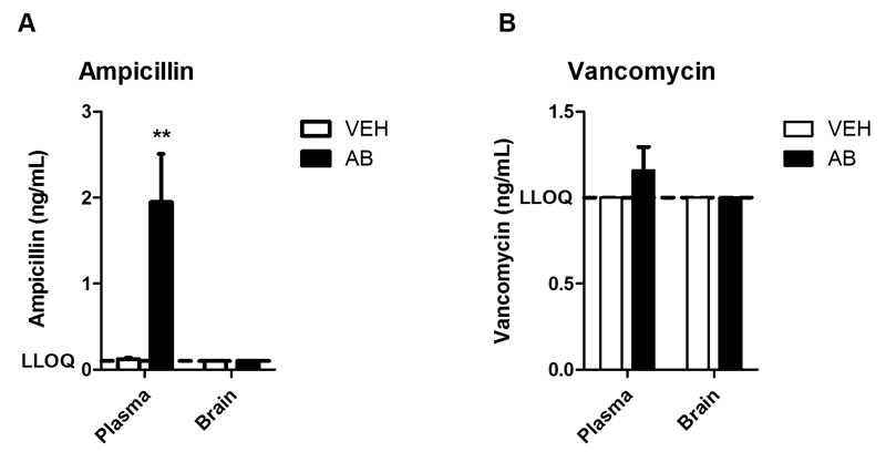
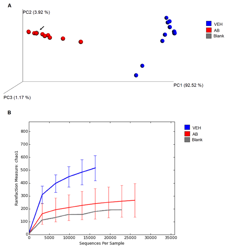
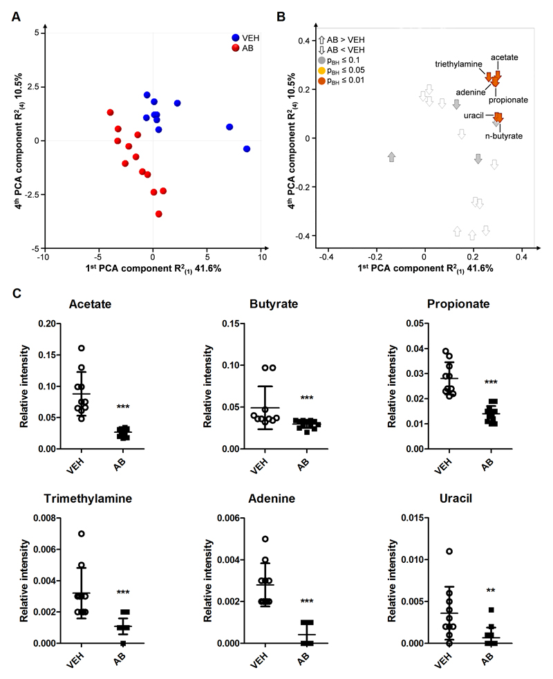
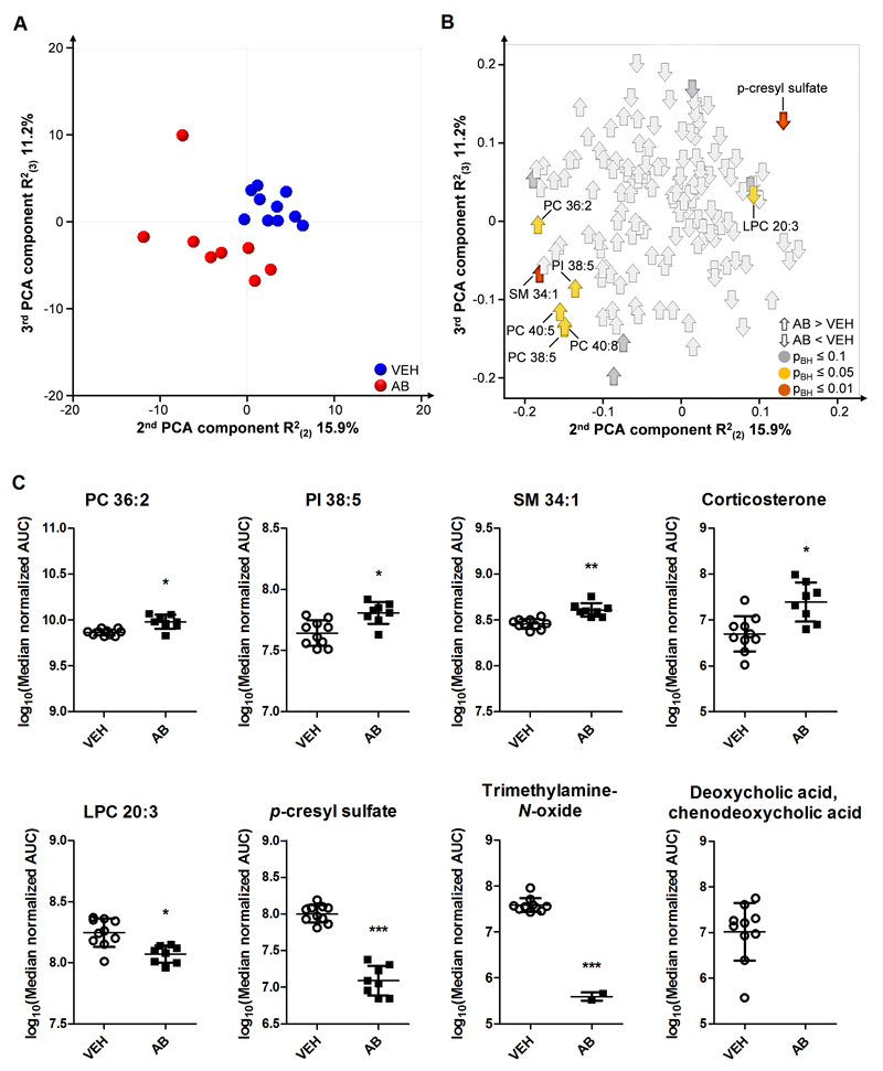
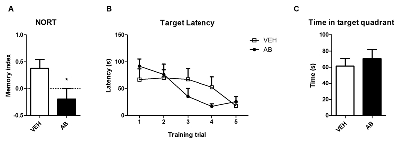
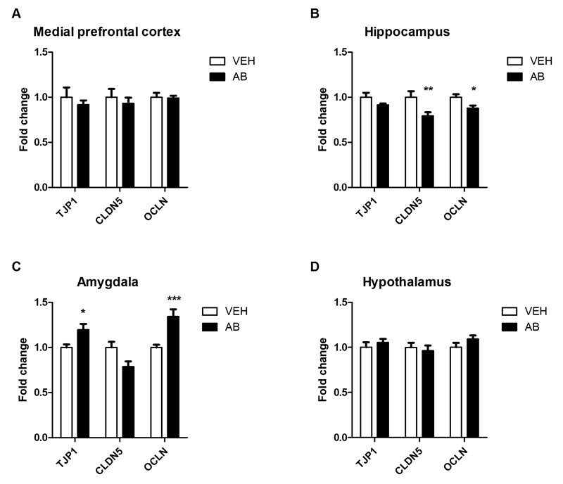
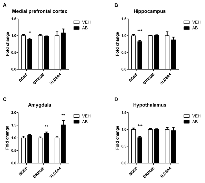
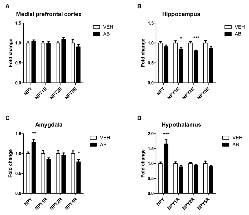
Emerging evidence indicates that disruption of the gut microbial community (dysbiosis) impairs mental health. Germ-free mice and antibiotic-induced gut dysbiosis are two approaches to establish causality in gut microbiota-brain relationships. However, both models have limitations, as germ-free mice display alterations in blood-brain barrier and brain ultrastructure and antibiotics may act directly on the brain. We hypothesized that the concerns related to antibiotic-induced gut dysbiosis can only adequately be addressed if the effect of intragastric treatment of adult mice with multiple antibiotics on (i) gut microbial community, (ii) metabolite profile in the colon, (iii) circulating metabolites, (iv) expression of neuronal signaling molecules in distinct brain areas and (v) cognitive behavior is systematically investigated. Of the antibiotics used (ampicillin, bacitracin, meropenem, neomycin, vancomycin), ampicillin had some oral bioavailability but did not enter the brain. 16S rDNA sequencing confirmed antibiotic-induced microbial community disruption, and metabolomics revealed that gut dysbiosis was associated with depletion of bacteria-derived metabolites in the colon and alterations of lipid species and converted microbe-derived molecules in the plasma. Importantly, novel object recognition, but not spatial, memory was impaired in antibiotic-treated mice. This cognitive deficit was associated with brain region-specific changes in the expression of cognition-relevant signaling molecules, notably brain-derived neurotrophic factor, N-methyl-d-aspartate receptor subunit 2B, serotonin transporter and neuropeptide Y system. We conclude that circulating metabolites and the cerebral neuropeptide Y system play an important role in the cognitive impairment and dysregulation of cerebral signaling molecules due to antibiotic-induced gut dysbiosis.

Keywords: Antibiotic; Brain; Cognition; Dysbiosis; GRIN2B; Gut; Metabolome; Microbiome; Neuropeptide Y; Serotonin transporter.


Figures

Fig. 1
Fig. 1. Experimental groups and time lines.
Mice of group A were subjected to a test battery including OFT, EPM test, TST and NORT. After sacrifice, tissues of group A animals were analyzed as indicated. Mice of group B were habituated and trained in the BM. Animals of group C were not subjected to any behavioral tests but euthanized on the day 10 (the day of cognitive evaluation in group A and B) to measure antibiotic concentrations in plasma and brain and investigate the effect of antibiotics alone on colon histology. In all experiments vehicle (VEH)- and antibiotic (AB)-treated animals were run in parallel, the number of mice in each group being indicated in brackets. OFT, open field test; EPM, elevated plus maze; TST, tail suspension test; NORT, novel object recognition test; BM, Barnes maze; qPCR, quantitative real-time polymerase chain reaction.
Fig. 2
Fig. 2. Brain concentrations of ampicillin and vancomycin are below the respective lower limit of quantification.
The graphs depict concentrations of ampicillin (A) and vancomycin (B) in plasma and brain after 10 days of antibiotic (AB) treatment. The bars represent means + SEM, n=6-8; **p ≤ 0.01 compared to vehicle (VEH)-treated mice, Mann-Whitney U test.
Fig. 3
Fig. 3. Antibiotic treatment strongly disrupts and diminishes the microbial community in the colon.
Mice were treated with antibiotic (AB) mix or vehicle (VEH) by gavage for 11 days. (A) Principal coordinate analysis (PCoA) plot based on weighted UniFrac distance between samples. The microbiome analysis blank is depicted as a dot highlighted by an arrow. Two individual dots of antibiotic-treated mice are not visible because they are overlaid by other dots. (B) Alpha-rarefaction curves using the chao1 index. Values in B represent median ± SD, n=10-12.
Fig. 4
Fig. 4. Distinct microbial metabolite levels in the colonic contents are markedly decreased by antibiotic treatment.
Mice were treated with antibiotic (AB) mix or vehicle (VEH) by gavage for 11 days. (A) Scores plot of the principal component analysis (PCA) with 21 identified colonic metabolites analyzed by 1H NMR, showing group separation in the first and fourth components. (B) Corresponding loadings plot, showing the contribution of metabolites to group separation. The metabolites are colored according to p-values calculated with the Mann-Whitney U test. (C) Graphs of significantly different metabolites. Vertical bars represent the group mean, whiskers SD, n=10-12; **p ≤ 0.01, ***p ≤ 0.001 compared to VEH-treated mice, Mann-Whitney U test.
Fig. 5
Fig. 5. Circulating metabolite levels are markedly altered by antibiotic treatment.
Mice were treated with antibiotic (AB) mix or vehicle (VEH) by gavage for 11 days. (A) Scores plot of the principal component analysis (PCA) with 148 identified circulating metabolites analyzed by LC-MS, showing group separation in the second and third components. (B) Corresponding loadings plot, showing the contribution of metabolites to group separation. Metabolites are colored according to Benjamini-Hochberg adjusted p-values. (C) Graphs of selected metabolites that were significantly different. Vertical bars represent the group mean, whiskers SD, n=8-10; *p ≤ 0.05, **p ≤ 0.01, ***p ≤ 0.001 compared to VEH-treated mice, Benjamini-Hochberg adjusted p-values. AUC, area under the curve; PC, phosphatidylcholine; PI, phosphatidylinositol; LPC, lysophosphatidylcholine; SM, sphingomyelin.
Fig. 6
Fig. 6. Object recognition memory is impaired in antibiotic-treated mice whereas spatial learning and memory remain intact.
Mice were treated with antibiotic (AB) mix or vehicle (VEH) by gavage for 11 days. In the NORT (A) a positive memory index (MI) indicates that the mice spent more time exploring the novel object than the known object. In the BM test (B,C) panel B shows the decrease in target latency with consecutive training trials, and panel C shows memorization of the target hole in the target quadrant on the probe day. Values represent means + SEM, n=6-8 (A), n=7-8 (B), n=6-7 (C); *p ≤ 0.05 compared to VEH-treated mice, t-test.
Fig. 7
Fig. 7. Antibiotic treatment alters tight junction protein mRNA expression in amygdala and hippocampus.
Mice were treated with antibiotic (AB) mix or vehicle (VEH) by gavage for 11 days. The panels show the expression of tight junction protein 1 (TJP1), claudin 5 (CLDN5) and occludin (OCLN) mRNA in the medial prefrontal cortex (A), hippocampus (B), amygdala (C), and hypothalamus (D). mRNA expression is expressed as fold change relative to VEH-treated mice. Values represent means + SEM, n=10-12; *p ≤ 0.05, **p ≤ 0.01, ***p ≤ 0.001 compared to VEH-treated mice, t-test.
Fig. 8
Fig. 8. Antibiotic treatment alters the expression of neural signaling-related molecules in a brain region-specific manner.
Mice were treated with antibiotic (AB) mix or vehicle (VEH) by gavage for 11 days. The panels show the expression of brain-derived neurotrophic factor (BDNF), N-methyl-D-aspartate receptor subunit 2B (GRIN2B) and serotonin transporter (SLC6A4) mRNA in the medial prefrontal cortex (A), hippocampus (B), amygdala (C), and hypothalamus (D). mRNA expression is expressed as fold change relative to VEH-treated mice. Values represent means + SEM, n=10-12; *p ≤ 0.05, **p ≤ 0.01, ***p ≤ 0.001 compared to VEH-treated mice, t-test.
Fig. 9
Fig. 9. Antibiotic treatment alters the expression of neuropeptide Y and its receptors in a region-specific manner.
Mice were treated with antibiotic (AB) mix or vehicle (VEH) by gavage for 11 days. The panels show the expression of neuropeptide Y (NPY) and the NPY receptors NPY1R, NPY2R and NPY5R in the medial prefrontal cortex (A), hippocampus (B), amygdala (C), and hypothalamus (D). mRNA expression is expressed as fold change relative to VEH-treated mice. Values represent means + SEM, n=10-12; *p ≤ 0.05, **p ≤ 0.01, ***p ≤ 0.001 compared to VEH-treated mice, t-test.

Comment in

References

    1. Antunes M, Biala G. The novel object recognition memory: neurobiology, test procedure, and its modifications. Cogn Process. 2012;13:93–110. doi: 10.1007/s10339-011-0430-z. - DOI - PMC - PubMed
    1. Attar A, Liu T, Chan WT, Hayes J, Nejad M, Lei K, Bitan G. A shortened Barnes maze protocol reveals memory deficits at 4-months of age in the triple-transgenic mouse model of Alzheimer's disease. PLoS One. 2013;8:e80355. doi: 10.1371/journal.pone.0080355. - DOI - PMC - PubMed
    1. Bajad SU, Lu W, Kimball EH, Yuan J, Peterson C, Rabinowitz JD. Separation and quantitation of water soluble cellular metabolites by hydrophilic interaction chromatography-tandem mass spectrometry. J Chromatogr A. 2006;1125:76–88. S0021-9673(06)00993-9 [pii] - PubMed
    1. Balderas I, Rodriguez-Ortiz CJ, Salgado-Tonda P, Chavez-Hurtado J, McGaugh JL, Bermudez-Rattoni F. The consolidation of object and context recognition memory involve different regions of the temporal lobe. Learn Mem. 2008;15:618–624. doi: 10.1101/lm.1028008. - DOI - PMC - PubMed
    1. Baldock PA, Lin S, Zhang L, Karl T, Shi Y, Driessler F, Zengin A, Hormer B, Lee NJ, Wong IP, Lin EJ, et al. Neuropeptide y attenuates stress-induced bone loss through suppression of noradrenaline circuits. J Bone Miner Res. 2014;29:2238–2249. doi: 10.1002/jbmr.2205. - DOI - PubMed

Substances